
Case Report
Ann Hematol Oncol. 2016; 3(2): 1076.
Spontaneous Resolution of Aleukemic Leukemia Cutis: A Case Report
Joseph S* and Liu Y
Department of Internal Medicine, Pinnacle Health Network, USA
*Corresponding author: Sarah Joseph, Department of Internal Medicine, Pinnacle Health Network, 111 S Front St, Third floor Brady Building: Harrisburg, PA, USA
Received: February 25, 2016; Accepted: February 25, 2016; Published: April 13, 2016
Abstract
A patient presented to her primary care physician with a skin rash; biopsy revealed leukemic cutis. Her peripheral blood demonstrated a slight increase in monocytes; however her bone marrow biopsy was negative for myeloblasts. The patient did not have any B symptoms and there was a complete resolution of her skin rash 3 months after diagnosis. The patient, 28 months after initial time of diagnosis, remained completely asymptomatic. This is the first reported case describing complete spontaneous resolution of leukemia cutis in an adult without a progression to systemic leukemia after more than 2 years of follow up.
Keywords: leukemia cutis; Spontaneous resolution
Introduction
Leukemia cutis is a nonspecific term to describe skin manifestations of leukemia [1]. It is defined as a cutaneous infiltration by malignant leukocytes and is often associated with progression to acute myeloid leukemia (AML), chronic myeloproliferative disease and chronic myelogenous leukemia [2]. An estimated 10-15% of patients with systemic AML develop LC [3]. Aleukemic leukemia cutis (ALC) is the manifestation of leukemic cells in the skin without infiltration in the peripheral blood and bone marrow and is often associated with poor outcome [4].
Case Presentation
An 85 year old Caucasian female was referred to our hematology oncology clinic because of a biopsy proven diagnosis of leukemia cutis. The patient has a past medical history significant for ER/PR positive breast cancerT1N0Mx, diagnosed 8 years prior. She underwent a left partial mastectomy, subsequently completed adjuvant radiation therapy, and took anastrozole for 5 years. In the summer of 2013, the patient noticed a rash on her right flank, described as a 6-10mm oval infiltrated pink plaque (Figure 1). She had no complaints except the skin rash. She denied any B-like symptoms. Her primary care doctor referred her to a dermatologist for a punch biopsy. Pathology reports revealed leukemia cutis with monocytic differentiation. Immunohistochemistry revealed cells diffusely positive for lysozyme and, to a lesser degree, CD68. Cells stained negative for CD3, CD45RO and myeloperoxidase. The biopsy results were confirmed by two institutions. Peripheral blood revealed an increase in monocytes. Bone marrow biopsy reported hyper cellular maturing trilineage hematopoiesis; there was no evidence of blast cells. The patient returned to the clinic 3 months later. She expressed no complaints and her peripheral blood at that time was unremarkable except for slight monocytosis. By November 2013, 6 months post diagnosis by skin biopsy, the rash had completely resolved (Figure 2). There was nothing left to re-biopsy, and the patient remained asymptomatic. Two years after the diagnosis of aleukemic leukemia cutis, there was no progression to systemic leukemia. The rash did not return and there was no evidence of active malignancy. Repeat bone marrow biopsy was deemed clinically unnecessary.
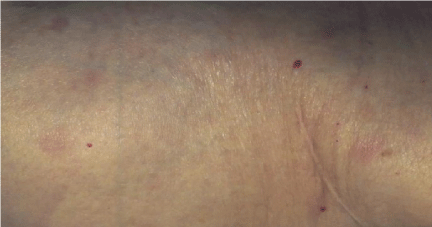
Figure 1: Leukemia Cutis.
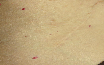
Figure 2: Spontaneous resolution of the rash.
Discussion
Aleukemic leukemia cutis was considered a dermatological curiosity until it led clinicians to an early diagnosis of acute myelogenic leukemia [5]. It is now thought to be a first sign of leukemia (Yonal I, 2011). In 1976, Yoder et al., demonstrated that the presence of ALC can lead to an early diagnosis of systemic leukemia, and described a pediatric case of ALC with subsequent bone marrow biopsy proven acute lymphoblastic leukemia [5]. In 1986, similar cases were reported of “aleukemic presentations” where both initial presentation and relapse of acute leukemia presented in the skin, with completely normal blood counts, but in which bone marrow biopsies led to the diagnosis of acute leukemia [6]. Our patient initially presented with a rash consistent with leukemic cutis, however both peripheral blood test and bone marrow biopsy failed to diagnose systemic leukemia. A complete resolution of aleukemia leukemia cutis has not been previously reported in the adult literature. In 2007, D’Orazio et al. described infantile leukemia cutis in a patient who presented with a single subcutaneous chloroma that resolvedover several weeks without any recurrence of disease [7]. However, to date there is a paucity of information on the pathophysiology of the spontaneous resolution of leukemic cutis in congenital leukemia. Furthermore, adult cases reported tend to characterize the presence of ALC with a subsequent diagnosis of systemic leukemia [4],[8],[9]. Our patient remained asymptomatic two years after follow up with a complete resolution of hercutaneous manifestations and no systemic progression. It is unclear if the patient’s own immune system is responsible of innately clearing the cutaneous infiltration of leukemic cells.
We present this case because, although aleukemic leukemia cutis has been previously described as being a precursor to the development of acute leukemias, some cases may not progress. The mechanism is still unclear and warrants further studies.
References
- Cho-Vega J, M. L. Leukemia Cutis. Am J Clin Pathol. 2008; 129: 130-142.
- Hussein M, A. b. Leukemia Cutis: Case Report and Review of Literature. Appl Immunohistochem Mol Morphol. 2010; 2: 190-191.
- Agis H, W. A. A comparative study on demographic, hematological, and cytogenetic findings and prognosis in acute myeloid leukemia with and without leukemia cutis. Ann Hematol. 2002; 81: 90-95.
- Benez A, M. A. Aleukemic Leukemia Cutis Presenting as Benign-appearing Exanthema. Acta Derm Venereol. 2001; 81: 45-47.
- Yoder F, S. R. Aleukemic Leukemia Cutis. Archives of Dermatology. 1979; 112: 367-369.
- Hansen R, B. J. Aleukemic Leukemia Cutis. Archives of Dermatology. 1986. 122: 812-814.
- D'Orazio J, P. J. Spontaneous Resolution of a Single Lesion of Myeloid Leukemia Cutis in an Infant: Case Report and Discussion. Pediatr Hematol Oncol. 2008; 25: 457-458.
- Vishalakshi V, T. R. Aleukemic Leukemia Cutis. Indian Journal of Dermatology, Venereology and Leprology. 2007; 73: 109-111.
- Martinez - Poventud G, F. J. Aleukemic leukemia cutis preceding acute monocytic leukemia: a case report. P R Health Sci J. 2008; 3: 256-258.