
Case Report
Ann Hematol Oncol. 2015; 2(9): 1063.
Urothelial and Renal Papillary Carcinomas Presenting as Paraneoplastic Florid Cutaneous Papillomatosis
Samhouri Y¹, Samudrala G¹ and Ban-Hoefen M²*
¹Department of Internal Medicine, Rochester Regional Health-Unity Hospital, USA
²Department of Interlakes Oncology and Hematology, University of Rochester, USA
*Corresponding author: Ban-Hoefen M, Department of Interlakes Oncology and Hematology, University of Rochester, 211 White Spruce Blvd, Rochester, NY 14623, USA
Received: October 01, 2015; Accepted: November 10,2015; Published: November 15, 2015
Abstract
Florid Cutaneous Papillomatosis (FCP) is a rare paraneoplastic syndrome similar to Malignant Acanthosis Nigricans and sign of Leser-Trélat. FCP is well associated with gastric adenocarcinoma. We report a rare case of FCP and malignant acanthosis nigricans as presenting signs that led to the diagnosis of three different malignancies, namely, urothelial cancer, renal papillary carcinoma and conjunctival carcinoma in-situ in a patient with Phosphatase Tensin (PTEN) germline mutation. FCP presents as eruptions of 1-3 mm diameter wart-like nodules on the hands, feet and face. Sudden onset of similar diffuse cutaneous lesions should elicit serious consideration for paraneoplastic disease and further workup to evaluate for an underlying cancer. This case adds to sparse literature on FCP and highlights their importance as paraneoplastic markers.
Keywords: Florid cutaneous papillomatosis; Renal papillary carcinoma; PTEN; Paraneoplastic
Abbreviations
FCP: Florid Cutaneous Papillomatosis; PTEN: Phosphatase and Tensin Homolog
Case Presentation
A 55 year old gentleman with hypertension, an excellent performance status and working full time as an electrical engineer, developed hematuria and diffuse skin eruptions over a month’s time. The patient has a very strong family history of malignancies. His Mother had breast cancer at age of 40, his father had bladder cancer at age of 70 and his paternal aunt had colon cancer. On physical exam, he had innumerable verrucous and filiform fleshy white hyperkeratotic papillomas over the face (Figure 1), neck and arms that spared the palms. Both conjunctivae were erythematous. Perianal skin was papillomatous with velvety hyper-pigmented area (Figure 2). The remainder of the physical exam was unremarkable. Complete blood count and chemistries were normal. CT scan of chest, abdomen and pelvis revealed enhancing lesions within the right collecting system and right retroperitoneal and aortocaval lymphadenopathy, in addition to a 4.8 cm mass in the upper pole of the left kidney (Figure 3). Biopsy of the three regions revealed right kidney invasive urothelial carcinoma, right retroperitoneal metastatic urothelial carcinoma and left kidney renal papillary neoplasm. The conjunctival biopsy showed carcinoma in-situ. He was started on neoadjuvant chemotherapy with Gemcitabine and Cisplatin. After two cycles of chemotherapy, there was significant improvement in the rash (Figure 4). After 4 cycles of chemotherapy, imaging revealed improvement in the number and dimensions of lymph nodes, stable size of the left renal mass, and shrinkage of the right collecting system mass. He subsequently underwent a right nephroureterectomy and retroperitoneal lymph node dissection. Unfortunately, the papillomas reappeared three months after the surgery. CT scan of the abdomen and pelvis showed new retroperitoneal lymphadenopathy. Core biopsy of these lymph nodes confirmed urothelial carcinoma.
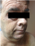
Figure 1:
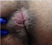
Figure 2:
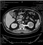
Figure 3:
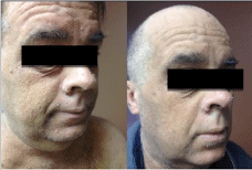
Figure 4:
Discussion
Florid Cutaneous Papillomatosis (FCP) is a rare condition [1], in which there are less than thirty cases reported in the literature so far. The onset of FCP often manifests before or concurrent with the diagnosis of internal malignancy. It is twice as common in men as in women with mean age of 58.5 years at diagnosis.
Most cases of FCP reported in the literature are associated with gastric adenocarcinoma [2,3]. Weger et al. in 2007 reported its association with lung and prostate cancers. In a review of 46 patients, Stieler and Plewig [4] noted that each patient had an underlying cancer, with more than half of the patients diagnosed with gastric adenocarcinoma. Other cancers that were reported include cancer of the urinary bladder, biliary ducts, ovaries, uterus, breast adenocarcinoma squamous cell carcinoma of the lungs, and non- Hodgkin lymphoma. Interestingly, our patient was simultaneously diagnosed with three different cancers, namely, urothelial carcinoma, renal cell papillary neoplasm and conjunctival carcinoma in-situ. This finding led to genetic testing which revealed PTEN germline mutation in the present case.
FCP is one of the cutaneous paraneoplastic syndromes, similar to malignant acanthosis nigricans, and the sign of Leser-Trélat. These should be viewed as part of a continuum because they can manifests together and share similar pathogenesis. Malignant acanthosis nigricans is clinically characterized by the appearance of thick velvety skin with hyper pigmentation located symmetrically in intertriginous areas. The sudden eruption of multiple seborrheic keratoses constituting the sign of Leser-Trélat may occur with malignant acanthosis nigricans and FCP. Each seborrheic keratosis is a discrete verrucous nodule that appears as if it has been stuck on the skin. These nodules should be easily distinguished from those of FCP. In this case, the patient was noted to have both FCP and malignant acanthosis nigricans as presenting findings of the malignancies, both of which resolved after surgical excision of his cancers and chemotherapy.
The pathophysiology of FCP is not well understood, Koyama et al suggested that the underlying malignancy induces FCP by secreting a factor similar to human epidermal growth factor or transforming growth factor-alpha that is transported in the blood to the skin surface and stimulate the development of keratinocytes by activating tyrosine kinase receptors in these cells, which in turn promote widespread mitotic and antiapoptotic cellular activity [5,6].
The main differential diagnosis of FCP is viral warts, which can be clinically indistinguishable from FCP. Although FCP can also present as eruptions of 1-3 mm diameter wart-like nodules on the hands, feet and face [7], there is no evidence of an underlying viral infection that is associated with viral warts. FCP differs histologically from viral warts by lacking epidermal vacuolization, parakeratosis and eosinophilic inclusions [8].
In one-third of cases, FCP heralds an underlying malignancy which improves after surgical resection of the cancer or chemotherapy treatment. Furthermore, the recurrence of FCP corresponds to cancer recurrence or progression [9], as seen in the present case. Topical 5-florouracil has demonstrated effectiveness as palliative treatment. Oral retinoids and smallpox vaccination proved to be beneficial systemic treatments as well.
Conclusion
This case adds to the sparse literature on Florid Cutaneous Papillomatosis (FCP) and highlights their importance as neoplastic markers. To our knowledge, this is the first case in literature describing FCP as a presenting sign of three different cancers in a patient with PTEN germline mutation. The sudden onset of similar diffuse cutaneous lesions should elicit serious consideration for paraneoplastic disease and further workup to evaluate for an underlying cancer.
References
- Schwartz RA. Skin cancer: Recognition and management. 2nd edition. Malden, MA: Blackwell. 2008; 283-304.
- Singhi MK, Gupta LK, Bansal M, Jain V, Kachhawa D. Florid cutaneous papillomatosis with adenocarcinoma of stomach in a 35 year old male. Indian J Dermatol Venereol Leprol. 2005; 71: 195-196.
- Yoo KH, Rho YK, Kim JY, Li K, Seo SJ, Hong CK. Cutaneous paraneoplastic syndrome in a patient with adenocarcinoma of unknown primary site syndrome. J Clin Oncol. 2009; 27: 309-311.
- Stieler W, Plewig G. [Acanthosis nigricans maligna and Leser-Trélat sign in double malignancy of the breast and stomach]. Z Hautkr. 1987; 62: 344-366.
- Schwartz RA. Florid cutaneous papillomatosis. Clin Dermatol. 1993; 11: 89-91.
- Koyama S, Ikeda K, Sato M, Shibahara K, Yuhara K, Fukutomi H, et al. Transforming growth factor-alpha (TGF alpha)-producing gastric carcinoma with acanthosis nigricans: An endocrine effect of TGF alpha in the pathogenesis of cutaneous paraneo- plastic syndrome and epithelial hyperplasia of the esophagus. J Gastroenterol. 1997; 32: 71-77.
- Mansouri P, Lotfi M, Naraghi M. Florid cutaneous papillomatosis, malignant acanthosis nigricans. Palmoplantar keratoderma, and gastric adenocarcinoma. Acta Med Iran. 1999; 37: 63-67.
- Jablonska S, Kozminska A, Chorzelski T. [An Unusual Variant of Acanthosis Nigricans. (Relations of Acanthosis Nigricans to Generalized Verrucosis)]. Dermatol Wochenschr. 1965; 151: 177-185.
- Schwartz RA, Burgess GH. Florid cutaneous papillomatosis. Arch Dermatol. 1978; 114: 1803-1806.