
Case Report
Ann Hematol Oncol. 2015;2(2): 1023.
Increased Incidence of T-Cell Malignancies in Patients with Chronic Lymphocytic Leukemia
Choi G1*, van den Broek EC3*, Stam OCG2, van Noesel CJM2, Tonino SH1 and Kater AP1
1Department of Hematology, University of Amsterdam, Netherlands
2Department of Pathology, University of Amsterdam, Netherlands
3Integraal Kankercentrum Zuid, Eindhoven, Netherlands
*Corresponding author: G Choi, University Medical Center Groningen, Department of Hematology (HPC DA21) Hanzeplein 1, 9713 GZ Groningen, The Netherlands
Received: December 04, 2014; Accepted: January 16, 2015; Published: January 19,2015
Abstract
We present a patient with chemotherapy-refractory Chronic Lymphocytic Leukemia (CLL) in whom postmortem examination showed hepatosplenomegaly, with both multiple small-cellular CLL lesions and large-cellular, monoclonal T-cell infiltrates. Following this case, the co-incidence of T-cell malignancies and CLL was studied using Dutch and American cancer registry databases. Analysis showed an excess risk for T-cell malignancies in CLL patients, with increased standardized incidence ratios compared with the general population and all cancer survivors in the databases. We hypothesize that CLL cells interact with T-cells in the microenvironment, facilitating malignant transformation.
Keywords: Chronic lymphocytic leukemia; T-cell lymphoma; Neoplastic cell transformation; Cohort studies
Introduction
Chronic Lymphocytic Leukemia (CLL) is a chronic lymphoproliferative disorder characterized by progressive accumulation of mature B-cells in peripheral blood and secondary lymphoid organs. Primary causes of death include infections and secondary malignancies [1], suggesting an immunodeficiency in CLL patients [2]. Recent epidemiological data demonstrated that the increased risk of other primary malignancies in CLL patients is independent of treatment or surveillance bias [3]. Most likely, CLL is not a disease limited to B-cells only, but also affects other components of the immune system due to extensive crosstalk between CLL cells and their microenvironment [4]. In this report, we present a patient with therapy-refractory CLL in whom post-mortem examination revealed the presence of a T-cell lymphoma. We further studied the coincidence of T-cell malignancies and CLL within two populationbased cancer registries, and found an excess risk of T-cell malignancies in patients with CLL.
Case Presentation
A 75-year-old male patient with therapy-refractory CLL was referred to our hospital. CLL had been diagnosed 7 years earlier (initially Rai 0) and during the last 3 years, he was intermittently treated with Chlorambucil. One month before admission, he had a painful cervical node, from which a biopsy revealed CD5 positive, cycline-D1 negative B-cells, compatible with CLL and without signs of transformation. Upon admission, patient presented with fever, dysphagia, and general malaise. His blood counts showed a pancytopenia with hemoglobin 7.9 g/dL (12.1 a month before), leukocytes 2,300/μl (50% lymphocytes), and platelets 10,000/μl (80,000 before). Without clinical signs of a specific infection, he was treated with broad-spectrum antibiotics. A bone marrow biopsy was performed, showing 80% CLL cells (positive for CD5/CD19/CD23/ CD20dim) and dysplasia in the myeloid and erythroid lineages. Within hours, he had progressive respiratory failure due to either sepsis or transfusion-related lung injury. Despite ventilatory support, patient’s condition rapidly deteriorated and he died within a few hours.
Post-mortem examination showed signs of multi-organ failure, including congested lungs without microbial infiltrates. There was hepatosplenomegaly and lymphadenopathy. Immunohistochemistry studies showed diffuse infiltrates of small, basophilic cells, positive for CD5, CD20 and CD23 (in liver, spleen, and bone marrow). Remarkably, the spleen contained, apart from CLL, areas with large cells with atypical nuclei positive for CD3, CD8, and Granzyme B, and negative for CD4, CD5, CD20, CD23, CD30, ALK1, CD79a, PAX-5, EBV, and keratin. Ki67 showed a high proliferation index in these areas. Altogether, this patient was found to have CLL and a high-grade peripheral T-cell lymphoma NOS (Figure 1). Molecular analyses of both spleen and liver lesions demonstrated clonal B-cell receptor and clonal T-cell receptor rearrangements, confirming both malignancies. Analysis was also performed on banked peripheral blood from 3 years earlier, excluding the presence of the malignant T-cell clone at an earlier stage of disease.
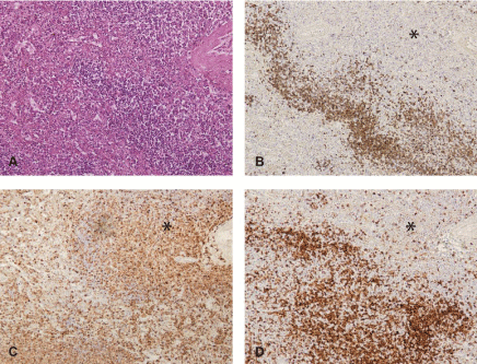
    Figure 1:  Spleen section with T-cell lymphoma (*) and CLL, stained
with (A) H&E, (B) CD79a, (C) CD3, (D) CD5 monoclonal antibodies (50x
magnification).
This case demonstrated that a large-cell T-cell lymphoma had developed in a patient with a chronic B-cell leukemia. In the classical Richter’s syndrome, CLL cells are believed to transform into a large cell B-cell lymphoma, after acquisition of additional molecular lesions [5]. Rarely, in large cellular lymphomas complicating CLL, there is any clonal relationship with CLL. There have been case reports of Hodgkin’s disease [6-8] and T-cell lymphoma [9-11] found in CLL patients.
Cohort study
To estimate the incidence of T-cell lymphomas among CLL patients, we performed a retrospective study within the Netherlands Cancer Registry (NCR) database. We retrieved all cancer cases diagnosed in the Netherlands between 1989 and 2008 and included patients diagnosed with a first primary invasive cancer (n =1,270,595). Patients were followed until the occurrence of T-cell lymphoma, death, or end of follow-up (December 31, 2008). The order of the cancer diagnosis was determined according to the International Rules for Multiple Primary Cancers (ICD-O, [12]). All T-cell malignancies diagnosed after CLL diagnosis were included, using ICD-O-2 codes 9700-9702, 9705, 9708, 9714, 9716-9719, 9827, 9831, 9834, 9948 and ICD-O-3 codes 9591, 9593, 9675, 9680, 9684, 9702-9704, 9713, 9823, 9825 combined with type=T-cell. As a measure of excess risk, Standardized Incidence Ratio (SIR) was calculated as the ratio of the observed number of cases in the cohort to the expected number of cases. The number of expected cases was calculated by multiplying the person-years at risk by the age-, gender-, and calendar year-specific incidence rates in the background population. 95% Confidence Intervals (95% CI) for SIRs were calculated based on a Poisson distribution. SAS software (SAS system 9.3, SAS Institute, Cary, NC) was used to perform the statistical analyses.
As shown in Table 1, Dutch CLL patients exhibited a threefold excess risk of developing a T-cell malignancy when compared with the entire Dutch population (SIR 3.0; 95% CI, 1.3-5.8). To correct for possible increased detection among cancer survivors, we also compared CLL patients with all Dutch cancer survivors. This yielded a SIR of 1.7, however statistically non-significant (95% CI, 0.71-3.3). As this might be due to the small size of the cohort, we performed the same analysis on the Surveillance, Epidemiology and End Results (SEER) database. We retrieved all registered cancer cases diagnosed in the United States (US) of America between 1973 and 2010 and included patients diagnosed with a first primary invasive cancer (n =6,316,679). End of follow-up date was December 31, 2010. We found that US CLL patients exhibited a more than two-fold risk forT-cell malignancies compared with all US cancer survivors (SIR 2.1; 95% CI, 1.7-2.7).
![]()
    Compared with


    CLL patients
    all cancers
    general population


    n
    Time at risk (PY)
    n (TCM)
    SIR
    95% CI
    SIR
    95% CI


    NCR
    13,504
    61,631
    8
    1.7
    (0.71-3.3)
    3.0
    (1.3-5.8)


    SEER
    90,721
    443,802
    72
    2.1
    (1.7-2.7)
    n/a
Table 1:  Increased rates of T-Cell Lymphoma (TCL) as a second primary
malignancy in patients with Chronic Lymphocytic Leukemia (CLL) as compared
with all cancer patients and the general population in the Netherlands (NCR) or
United States of America (SEER). CI, confidence interval; n, number of patients;
n/a, not available; PY, patient-years; SIR, standardized-incidence ratio.
Discussion
We here presented a patient with therapy-refractory CLL who died from multi-organ failure, in whom a T-cell lymphoma was detected at post-mortem examination. Within the Dutch NCR and larger US SEER databases, the epidemiological data corroborate the hypothesis that CLL predisposes patients for the development of T-cell malignancies. Indeed, there is a biologic rationale for increased incidence of clonal T-cell disorders in CLL. In CLL, there is an expansion of T-cells with a globally impaired function characterized by a pseudo-exhausted phenotype [13]. Also, microarray and functional studies showed that T-cells from CLL patients undergo significant changes on a molecular and structural level [14]. Clearly, CLL cells and T-cells have reciprocal interactions, inducing both expansion and intrinsic changes, and possibly facilitating malignant transformation. Of note, other factors may play a role in the pathogenesis of T-cell malignancies in CLL patients. We cannot exclude that previous treatment and/or immunosuppression would contribute to the pathogenesis of T-cell malignancies. In previous case reports, treatment with Chlorambucil, Fludarabine, and various immunosuppressive agents were described [10,11]. Compared with untreated follicular lymphoma patients, untreated CLL patients have a two-fold increased risk of second malignancies, as shown in a large Canadian cohort study [3]. Prolonged Chlorambucil treatment was not associated with an increase in second malignancies in a cohort of CLL patients [15], whereas most recently Benjamini et al. demonstrated that frontline treatment with Fludarabine, Cyclophosphamide, and Rituximab was associated with an increased risk of therapy related acute myeloid leukemia and Richter’s syndrome [16]. In these cohort studies, no cases of T-cell malignancy were described.
In conclusion, albeit the low incidence, CLL patients have an increased risk of developing T-cell malignancies. It remains to be established whether malignant transformation of T-cells is facilitated by interactions with CLL cells, direct mutations induced by chemotherapy, or deficiency in adequate immunosurveillance related to CLL and/or use of immunomodulating agents.
- Wierda WG, O'Brien S, Wang X, Faderl S, Ferrajoli A, Do KA, Garcia-Manero G . Characteristics associated with important clinical end points in patients with chronic lymphocytic leukemia at initial treatment. J Clin Oncol. 2009; 27: 1637-1643.
- Molica S. Second neoplasms in chronic lymphocytic leukemia: incidence and pathogenesis with emphasis on the role of different therapies. Leuk Lymphoma. 2005; 46: 49-54.
- Beiggi S, Johnston JB, Seftel MD, Pitz MW, Kumar R, Banerji V, et al. Increased risk of second malignancies in chronic lymphocytic leukaemia patients as compared with follicular lymphoma patients: a Canadian population-based study. Br J Cancer. 2013; 109: 1287–1290.
- Burger JA. Nurture versus nature: the microenvironment in chronic lymphocytic leukemia. Hematology Am Soc Hematol Educ Program. 2011; 2011: 96-103.
- Rossi D, Gaidano G. Richter syndrome: molecular insights and clinical perspectives. Hematol Oncol. 2009; 27: 1-10.
- Brecher M, Banks PM. Hodgkin's disease variant of Richter's syndrome. Report of eight cases. Am J Clin Pathol. 1990; 93: 333-339.
- Fayad L, Robertson LE, O'Brien S, Manning JT, Wright S, Hagemeister F, et al. Hodgkin's disease variant of Richter's syndrome: experience at a single institution. Leuk Lymphoma. 1996; 23: 333-337.
- Weisenberg E, Anastasi J, Adeyanju M, Variakojis D, Vardiman JW. Hodgkin's disease associated with chronic lymphocytic leukemia. Eight additional cases, including two of the nodular lymphocyte predominant type. Am J Clin Pathol. 1995; 103: 479-484.
- Lee A, Skelly ME, Kingma DW, Medeiros LJ. B-cell chronic lymphocytic leukemia followed by high grade T-cell lymphoma. An unusual variant of Richter's syndrome. Am J Clin Pathol. 1995; 103: 348-352.
- Martinez A, Pittaluga S, Villamor N, Colomer D, Rozman M, Raffeld M, et al. Clonal T-cell populations and increased risk for cytotoxic T-cell lymphomas in B-CLL patients: clinicopathologic observations and molecular analysis. Am J Surg Pathol. 2004; 28: 849-858.
- Novogrudsky A, Amorosi EL, Gottesman SR. High-grade T-cell lymphoma complicating B-cell chronic lymphocytic leukemia: an unusual manifestation of "Richter's syndrome". Am J Hematol. 2001; 66: 203-206.
- Working Group Report. International rules for multiple primary cancers (ICD-0 third edition). Eur J Cancer Prev. 2005; 14: 307-308.
- Riches JC, Davies JK, McClanahan F, Fatah R, Iqbal S, Agrawal S, et al. T cells from CLL patients exhibit features of T-cell exhaustion but retain capacity for cytokine production. Blood. 2013; 121: 1612-1621.
- Görgün G, Holderried TA, Zahrieh D, Neuberg D, Gribben JG. Chronic lymphocytic leukemia cells induce changes in gene expression of CD4 and CD8 T cells. J Clin Invest. 2005; 115: 1797-1805.
- Callea V, Brugiatelli M, Stelitano C, Gentile M, Nobile F, Morabito F. Incidence of second neoplasia in patients with B-cell chronic lymphocytic leukemia treated with chlorambucil maintenance chemotherapy. Leuk Lymphoma. 2006; 47: 2314-2320.
- Benjamini O, Jain P, Trinh L, Qiao W, Strom SS, Lerner S, et al. Second cancers in patients with chronic lymphocytic leukemia who received frontline fludarabine, cyclophosphamide and rituximab therapy: distribution and clinical outcomes. Leuk Lymphoma. Informa Clin Med. 2014; 1–8.