Research Article
Ann Hematol Oncol. 2025; 12(2): 1477.
Bone Marrow Metastasis of Rhabdomyosarcoma Mimicking Acute Leukemia: A Case Report
Ameur O1,2*, Tarbi M1,2, Bernoussi M1,2, Orchi I1,2, Essahli K1,2 and Zahid H1,2
1Department of hematology, Mohammed V Military Training Hospital, Rabat, Morocco
2Faculty of Medicine and Pharmacy, University Mohammed V, Rabat, Morocco
*Corresponding author: Ameur Othmane, Department of hematology, Mohammed V Military Instruction Hospital, Rabat, Morocco Email: othmane_ameur@um5.ac.ma
Received: March 24, 2025 Accepted: April 11, 2025 Published: April 15, 2025
Abstract
Introduction: Infiltration of the bone marrow by non-hematopoietic malignant tumors poses a diagnostic challenge due to clinical manifestations resembling leukemia. We present a case of orbito-palpebral rhabdomyosarcoma (RMS) with bone marrow metastasis, mimicking acute leukemia both clinically and morphologically.
Case Report: A 21-year-old female with no significant medical history presented with orbito-palpebral RMS. Following chemotherapy, she developed anemia requiring hospitalization for further evaluation. Blood analysis revealed bicytopenia, and a bone marrow smear showed complete megakaryocytedepleted marrow infiltration by blast-like cells, initially suggestive of acute leukemia. However, histopathological analysis of the osteomedullary biopsy confirmed metastatic RMS infiltration, with tumor cells exhibiting characteristic immunohistochemical markers.
Discussion: RMS is the most common soft tissue sarcoma in children and adolescents, typically localized to the head, neck, urogenital tract, and extremities. Metastatic spread to organs such as the bone marrow is rare, and its morphological similarity to acute leukemia may lead to misdiagnosis. Bone marrow involvement is often associated with advanced disease and poor prognosis.
Conclusion: Bone marrow metastasis of RMS should be considered in patients presenting with cytopenias and atypical morphological features to avoid unnecessary treatments. Accurate diagnosis relies on histopathological confirmation, particularly when leukemia-like features are observed. This case highlights the critical role of interdisciplinary collaboration (hematology, pathology, oncology) in managing rare presentations of sarcomas.
Keywords: Rhabdomyosarcoma; Bone marrow metastases; Acute leukemia mimicry; Immunohistochemical markers (Myogenin/Desmin)
Introduction
Infiltration of the bone marrow by non-hematopoietic malignant tumors is a diagnostic challenge due to clinical manifestations resembling to acute leukemia. These patients are often managed for aberrant hematologic disorders, leading to delayed diagnosis.
Rhabdomyosarcoma (RMS), though rare, is a common childhood cancer and the most frequent soft tissue sarcoma in children. The overall incidence in individuals under 20 years is 4.5 cases per million population [1]. In the United States, approximately 350 new cases are reported annually. According to the Surveillance Epidemiology and End Results (SEER) program, RMS incidence varies by age and histopathology [1]. We present a case of orbito-palpebral RMS with bone marrow metastases, clinically and morphologically mimicking acute leukemia.
Objective
To highlight bone marrow metastases of RMS as a differential diagnosis in patients presenting with acute leukemia-like symptoms and atypical marrow abnormalities.
Case Presentation
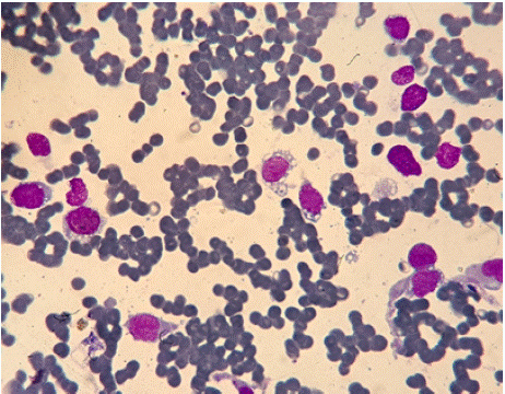
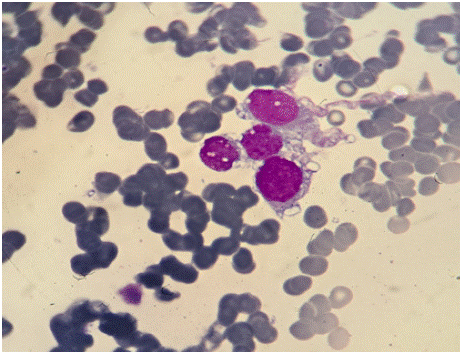
A 21-year-old female with no significant medical history presented with a progressively enlarging right orbito-palpebral mass. Imaging revealed a homogeneous orbital lesion. Biopsy with histopathological analysis suggested rhabdomyosarcoma. Post-treatment follow-up revealed the onset of anemia, prompting hospitalization for further evaluation. Blood count showed bicytopenia, and bone marrow smear demonstrated complete megakaryocyte-depleted marrow infiltration by blast-like cells, occasionally forming syncytial clusters (Figure 1-2), raising suspicion of post-chemotherapy acute leukemia.

    Figure 1: Bone marrow smear (MGG stain; 40x magnification) showing
diffuse infiltration by numerous blast-like cells.

    Figure 2: Bone marrow smear (MGG stain; 100x magnification) highlighting
cells with basophilic vacuolated cytoplasm, irregular borders, loose
chromatin, high nuclear-to-cytoplasmic ratio, and syncytial clustering.
However, bone marrow infiltration by metastatic RMS cells was confirmed via osteomedullary biopsy, with tumor cells showing nuclear positivity for Myogenin and Desmin on immunohistochemistry.
Discussion
RMS is the most common soft tissue sarcoma in children and adolescents, typically localized to the head, neck, urogenital tract, and extremities. While local complications of primary sarcomas cause significant morbidity, the most severe outcomes arise from hematogenous dissemination. For most soft tissue malignancies, the lungs are the primary metastatic site, followed by bone marrow, liver, breasts, and brain [2]. Approximately 20% of RMS patients present with metastases at diagnosis [3]. However, isolated bone marrow involvement as the sole site of progression or recurrence is rare, particularly without evidence of a primary tumor [4]. The morphological and clinical resemblance of metastatic RMS to acute leukemia may lead to misdiagnosis [5].
Bone marrow involvement is typically a poor prognostic indicator, often associated with extensive multifocal metastatic disease [6]. When bone marrow infiltration is the initial manifestation, symptoms are nonspecific: bone/joint pain, fever, nausea, vomiting, lethargy, anorexia, anemia, or hypercalcemia [4].
A PubMed review identified 42 reported cases of RMS with leukemia-like presentations, the earliest dating back four decades (Table 1) [5].
    Category
    Number of Cases (n=42)


    RMS with    leukemia-like onset
    39


    Known RMS    with late metastases
    2


    Primary site unidentified
    1


    Misdiagnosed as ALL*
    3


    Misdiagnosed as AML**
    2


    *ALL: Acute Lymphoblastic Leukemia; **AML: Acute Myeloid Leukemia.
Table 1: Analysis of 42 Reported Cases of RMS with Leukemia-like
Presentation.
In our case, bone marrow metastases of RMS displayed as blastlike cells, mimicking acute leukemia. Diagnosis was confirmed via Myogenin and Desmin positivity on bone marrow biopsy.
Conclusion and Perspectives
Bone marrow metastases of RMS should be considered in patients with cytopenias and atypical morphological features to avoid unnecessary treatments. This highlights the critical role of interdisciplinary collaboration (hematology, pathology, oncology) in managing rare presentations of sarcomas.
Consent
The patient gave informed consent for this publication after explaining the implications and the importance of the study.
Ethical Approval
According to Articles 2 and 3 of the Internal Regulations of the Biomedical Research Ethics Committee (CERB), this article is exempt from the Ethics Committee's approval.
References
- Zhen H, Liu Z, Guan H, Ma J, Wang W, Shen J, et al. Second Malignant Neoplasms in Patients with Rhabdomyosarcoma. Front Oncol. 2021; 11: 1–6.
- Shapeero LG, Couanet D, Vanel D, Ackerman L V., Terrier-Lacombe MJ, Flamant F, et al. Bone metastases as the presenting manifestation of rhabdomyosarcoma in childhood. Skeletal Radiol [Internet]. 1993; 22: 433– 438.
- Lee DH, Park CJ, Jang S, Cho YU, Seo JJ, Im HJ, et al. Clinical and Cytogenetic Profiles of Rhabdomyosarcoma with Bone Marrow Involvement in Korean Children: A 15-Year Single-Institution Experience. Ann Lab Med [Internet]. 2018; 38: 132–138.
- Shinkoda Y, Nagatoshi Y, Fukano R, Nishiyama K, Okamura J. Rhabdomyosarcoma masquerading as acute leukemia. Pediatr Blood Cancer [Internet]. 2009; 52: 286–287.
- López-Andrade B, Duran MA, Torres L, García-Recio M, Lo Riso L, Formica A, et al. Rhabdomyosarcoma debut masquerading as acute lymphoblastic leukemia: A case report and review of the literature. Clin case reports [Internet]. 2019; 7: 1545–1548.
- Etcubanas E, Peiper S, Stass S, Green A. Rhabdomyosarcoma, presenting as disseminated malignancy from an unknown primary site: a retrospective study of ten pediatric cases. Med Pediatr Oncol [Internet]. 1989; 17: 39–44.