
Case Report
Austin Gynecol Case Rep. 2023; 8(3): 1046.
Mitotically Active Cellular Fibroma of the Ovary: A Case Report
Vesna Antovska; Eva Sozovska Belchovska*; Iva Malahova Gjoreska; Angela Chipurovska; Ivona Dimovska
Department of Gynecology and Obstetrics, University of Clinical Gynecology and Obstetrics, Macedonia
*Corresponding author: Eva Sozovska Belchovska Department of Gynecology and Obstetrics, University of Clinical Gynecology and Obstetrics, Macedonia. Email: esozovska@yahoo.com
Received: October 02, 2023 Accepted: October 30, 2023 Published: November 06, 2023
Abstract
Ovarian fibromas are benign sex cord stromal tumors composed of fibroblastic cells within a collagenous stroma. Fibromas are composed of spindled, ovoid to round cells and collagenous stroma. There are three types of benign fibromas: Fibroma with minor sex cord elements, cellular fibroma and mitotically active cellular fibroma. Fibromas are most common ovarian stromal tumors and encompasses approximately 4% of all ovarian tumors. Cellular fibromas represent approximately 10% of all ovarian fibromas. According to published studies median age of patients with ovarian fibroma is 48 years and are uncommon before 30. The potential of mitotically active cellular fibromas for reccurence and malignant transformation is low (approximately 1%) but possible. So, diagnosis od MACF is important in order to avoid misdiagnosis with ovarian fibrosarcomas and to avoid unnecessary overtreatment. Also, because of low but possible malignant potential in case of MACF, long term follow-up should be recommended.
We report a case of mitotically active cellular fibroma in a 68 year old woman with previous vaginal hysterectomy without adnexectomy who applied to our clinic with mild pelvic pain and discomfort. Our intention is to point out its rarity among ovarian tumors and need for long term follow-up, because of its low but possible potential for reccurence and malignant transformation.
Case Report
A 68 year old woman presented to our clinic with mild pelvic pain and discomfort. Four and a half years previously, she had undergone total vaginal hysterectomy without adnexectomy because of uterine prolapse. On arrival she was in a relatively good condition, only with mild subjective symptoms like mild pelvic pain and discomfort.
In her medical history there was colonoscopy with resection of a benign colon polyp before 1 year. Of comorbidities she had arterial hypertension. In her gynecological history there was total vaginal hysterectomy without adnexectomy due to uterine prolapse, and after operation she didn’t have routine gynecological examinations beside recommendations. Bimanual vaginal examination revealed solid timorous formation, left and behind vaginal vault with size approximately like woman’s fist. Laboratory test showed normal values. Tumor markers (CEA,Ca 19-9, Ca 72-4, Ca 125, Ca 15-3) were in normal ranges. On transvaginal sonography, a solid left adnexal mass was noted, measuring 56mm and according to sonography features in favor of ovarian fibroma.
After complete evaluation and preparation the patient underwent operation. Laparotomy was done with low abdominal transverse approach (sec.Pfannenstiel). During operation there was detected left adnexal mass with smooth surface, solid and cystic parts and size like a woman’s fist. Also there were multiple adhesions between adnexal mass and sigmoid colon. There was no ascites. Subsequently extensive adhesiolysis was performed and after that left adnexa with adnexal mass were excised without any difficulty. Contralateral ovary seemed normal but due to age of the patient right adnexectomy was performed. A small part of omentum was also excised for histopathologic evaluation. Operative material was sent for histopathologic evaluation. Also peritoneal fluid obtained by peritoneal washing was sent for cytological examination. Additionally on previously detected vaginal condylomas was performed biopsy.
Final pathology result was reported as: Cellular fibroma of the left ovary, Chronic billateral salpingitis and Vaginal condyloma acuminata.
In histopathological report was described that operative material consists of: Left adnexa weighing 128gr. Left ovary with dimensions 8,5×6,5×4,5cm with lobulated surface and grey-yellow colour. On dissection there was uniform grey-white parenchime with small cystic areas in the central part of the mass.
Additionally histopathologic evaluation has been enhanced by immunohistochemical staining with: ki67,SMA, CD10,ER and PR.
Other operative materials were: Right adnexa, small piece of omentum with regular macromorphological structure and specimens obtained by vaginal biopsy of vaginal condylomas.
Microscopically, specimens of left ovary were consisted of elongated smooth muscle cells in spindeled and fascicular arrangement and without nuclear atypia but enhanced cellularity. Between cells were deposites of collagen. There were detected 5 mitosis in 10 High Power Fields (HPFs). Tumor cells displayed positivity for SMA, PR, and focally to 5% for ER and ki67. On specimen of uterine tubes was identified inflammatory infiltration. On other specimens there were no microscopically detected abnormalities. Specimens of vaginal biopsy confirmed vaginal condyloma acuminata. So,according to number of mitoses per 10HPFs , the final diagnosis was confirmed as mitotically active cellular fibroma of ovary.
There were no post-operative complications and she was discharged in good condition with given advice for regular follow-up.
Discussion
Sex-cord stromal tumors are second most common ovarian tumors after epithelial ovarian tumors. Ovarian fibromas are the most common tumors in the group of SCST. Ovarian fibromas are benign stromal tumors composed of fibroblastic cells within a variably collagenous stroma [3]. Fibromas encompasses approximately 4% of all ovarian tumors, and cellular fibromas represent approximately 10% of all ovarian fibromas [3]. Most common in perimenopausal and menopausal women, average: 48, <10% younger than 30 [2]. Etiology is unknown but it is thought that the main cause for fibroma formation is: neoplastic transformation of ovarian stromal cells due to hereditary or sporadic genetic abnormalities [3]. Fibromas are most commonly asymptomatic. Most fibromas are found incidentally during pelvic or sonographic examination [1]. Less common symptoms and signs are abdominal distention and pelvic discomfort, abdominal pain ( usually due to ovarian torsion) , urinary frequency , ascites and right hydrothorax associated with Meig’s syndrome, rarely estrogen secretion (when theca cells are dominant), nonspecific increase in serum Ca125 [3] and other minor signs and symptoms. Average size of fibroma is 6cm [2]. According to Irving et al., 2006, the mean tumor size of cellular fibromas was 8.0 cm and 9,4cm for MACFs [6]. Bilateral masses in <10% of patients [2]. Under age 30 often are associated with Gorlin’s syndrome [3]. Gorlin’s syndrome is associated with early basal cell carcinomas, keratosis of the jaw, calcification of the dura, mesenteric cysts and bilateral ovarian fibromas [2]. In preoperative evaluation , ultrasonography and computed tomography are helpful diagnostic methods. Preoperative evaluation (computed tomography) for possible lymph node enlargement or intraabdominal spread is indicated for patients in whom malignancy is a significant possibility [2]. Final diagnosis is established by histologic evaluation [2,3]. Ultrasonographically fibroma is solid, homogeneous mass with posterior acoustic shadowing,similar to a pedunculated subserosal uterine leiomyoma [3] (AJR Am J Roentgenol 1985;144:1239). Also it may have heterogeneous echogenicity especially in tumors with necrosis, hemorrhage or cystic degeneration [3] (Figure 1).
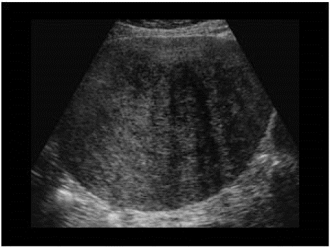
Figure 1: Ultrasound image of ovarian fibroma.
Grossly fibromas are described as well circumscribed masses with smooth, lobulated, soft and grey-yellow surface with possible cystic degeneration and calcifications [3]. Usually are unilateral. Pathologic findings include fibroblasts and spindle cells with intercellular collagen. Occasional mitoses are usually up to 3 mitosis per 10 high power fields [3]. Cellular fibromas have densely cellular pattern with little intercellular collagen [3] and up to 3 mitosis per 10HPFs. “Mitotically active cellular fibroma”has been proposed for cellular fibroma with =4 mitosis per 10HPFs [3] (Am J Surg Pathol 2006;30:929).
According to literature fibroblastic tumors are generally divided on fibromas and fibrosarcomas. The difference between them is in the number of mitosis at 10HPFs. Cellular fibromas have =4 mitosis per 10HPFs, have normal nucleus and benign feature. Otherwise malignant fibroblastic tumors called fibrosarcomas have more than 4 mitosis per 10HPFs and have nuclear atypia. Additionaly there are rare cases of fibroblastic tumors that have more than 4 mitosis per 10HPFs but benign features and are called “mitotically active cellular fibromas”.
Management of fibroblastic tumors depends of their appearance, age of the patient and fertility- sparing desire. Generally surgical exploration and resection is adequate [2]. In older women hysterectomy with bilateral adnexectomy is generally performed. Malignant fibromas are rare and treatment is more specific.
Prognostic factors for cellular fibromas are excellent in most cases, but ovarian surface involvement and extraovarian adhesions occur in 6% of cellular fibromas and 10% of mitotically active cellular fibromas [3] (Am J Surg Pathol 2006;30:929). Extraovarian spread at surgery is 11% of cellular fibromas and 13% of mitotically active cellular fibromas [3] (Am J Surg Pathol 2006; 30: 929). Cellular fibromas may recur, often after a long interval, warranting long term follow up [3] (Cancer 1981;47:2663). Especially, long term follow up is recommended for cellular fibromas associated with rupture and adhesions due to increased risk of recurrence [3].
Conclusion
Getting clear pathologic diagnosis of the fibroma is significant. It’s very important to differentiate whether it is benign or malignant fibroblastic tumor. Presence of =4 mitosis per 10 HPFs is not sufficient to diagnose fibrosarcoma in the absence of significant cytologic atypia and that is very important in order to avoid unnecessary overtreatment. Mitotically active cellular fibromas are rare tumors but their detection is important because of their very low but possible potential for recurrence (especially in presence of adhesions) and malignant transformation. So, mitotically active cellular fibromas need more close follow-up in contrast to benign fibromas with up to 3 mitosis per 10HPFs.
References
- Hoffman BL, Schorge JO, Bradshaw KD, Halvorson LM, Schaffer JI, Corton MM, et al. Gynecology. 3rd ed. 2016; 767-75.
- Smith RP. Netter’s obstetrics & gynecology. 2nd ed. 2009; 341-2.
- Gulisa Turashvili MD, et al. Ovary. Sex cord stromal tumors; 2021. Fibroma. PathologyOutlines.com. Avaliable at. Available from: http://www.pathologyoutlines.com/topic/ovarytumorfibroma.
- Yildirim N et al. Mitotically active cellular fibroma of the ovary:a case report. J Turk Soc Obstet Gynecol. 2015; 1: 53-5.
- Zong L, Lin M, Fan X. Mitotically active cellular fibroma of ovary should be differentiated from fibrosarcoma: a case report and review of literature. Int J Clin Exp Pathol. 2014; 7: 7578-82.
- Irving JA, Alkushi A, Young RH, Clement PB. Cellular fibromas of the ovary: a study of 75 cases including 40 mitotically active tumors emphasizing their distinction from fibrosarcoma. Am J Surg Pathol. 2006; 30: 929-38.