
Case Report
Austin Gynecol Case Rep. 2023; 8(1): 1036.
Postpartum Juvenile Granulosa Cell Tumor: A Case Report
V Antovska¹, D Dabeski¹, IA Papestiev¹, MP Ilieva¹, EA Kamberi¹, ES Belchovska¹* and P Zdravkovski²
¹University Clinic of Gynecology and Obstetrics, Faculty of Medicine – UKIM, Republic of North Macedonia
²Institute of pathology, Faculty of Medicine – UKIM, Republic of North Macedonia
*Corresponding author: Eva Sozovska Belchovska University Clinic of Gynecology and Obstetrics, Faculty of Medicine – UKIM, Skopje, Republic of North Macedonia
Received: December 06, 2022; Accepted: January 18, 2023; Published: January 25, 2023
Abstract
Granulosa Cell Tumors (GCTs) are extremely rare, sex cord-stromal tumors constituting only 1% to 2% of all ovarian malignancies. On the basis of age of onset and pathohistological characteristics, these tumors are subdivided into two distinct forms, the adult type (AGCT) and the juvenile type (JGCT), representing 95% and 5% of the tumors, respectively. Compared to the adult type, which is more common in the fifth decade, JGCT is rarely seen and the majority (90%) is reported in prepubertal individuals or those aged less than 30 years. We report an interesting case of a 24-years old woman with enormous ovarian juvenile granulosa cell tumor of the right ovary. Considering all the anamnestic data and ultrasound reports before and during the first pregnancy, where there has been no evidence for the presence of any tumor, we came to conclusion that the tumor grew rapidly during the first year since delivery. Maybe the tumor was present during the pregnancy or even before, but most probably it was not noticed because it small initial size or it was masked by the growing uterus at ultrasound. The first data for the tumor existence is 3 months before the surgical treatment.
Keywords: Ovarian juvenile granulosa cell tumor; Postpartal period
Abbreviations: Hgb: Hemoglobin; RBC: Red Blood Cells; Hct: Hematocrit; GCTs: Granulosa Cell Tumors; JGCT: Juvenile Granulosa Cell Tumor; AGCL: Adult Granulosa Cell Tumor
Introduction
Granulosa Cell Tumors (GCTs) are extremely rare, sex cord-stromal tumors constituting only 1% to 2% of all ovarian malignancies. On the basis of age of onset and pathohistological characteristics these tumors are subdivided into two distinct forms, the adult type (AGCT) and the juvenile type (JGCT), representing 95% and 5% of the tumors, respectively. Compared to the adult type, which is more common during the fifth decade, JGCT is rarely seen and the majority (90%) are reported during puberty or in patients younger than 30 years of age [1].
Clinically, abdominal pain and abdominal distention are common symptoms [1]. Although the majority of JGCT patients are easily diagnosed on the basis of these typical clinical symptoms in the early stage (Stage I) and usually have a benign clinical course after unilateral salpingo-oophorectomy, small portion of patients are diagnosed in more advanced stages (stage II-IV) and usually do not have typical clinical symptoms and clinical course is with unfavorable outcome [1].
Case History
We present a case of a 24-year-old woman, with abdominal distension and abdominal pain on palpation, and with absence of menstrual cycle for 6 months.
Our patient delivered 18 months before the diagnosis of this ovarian mass. She delivered vaginally, in full term, without any complications. During pregnancy, there were no symptoms, signs or any pelvic tumors described on regular ultrasonography exams. During the years before pregnancy, our patient has been treated for acne vulgaris and alopecia areata by a dermatologist with local therapy. There is no hormonal status or any gynecological ultrasonography exams dating from this period that could indicate the existence of ovarian mass even during this period of time, before the pregnancy.
After delivery, our patient has been breastfeeding her child during the period of seven months, when she stopped to breastfeed because of the low milk supply. After suspending the breastfeeding, she only had two regular menstrual cycles after which the period of amenorrhea has occurred.
Seven months before the surgery, the patient had noticed her abdomen distending and growing rapidly. First data of the existence of pelvic mass were collected three months before the surgery. A large pelvic mass was diagnosed by ultrasonography, which probably originated from right ovary with diameter 12x11cm. After this first exam, the patient became SARS-CoV-2 + which delayed further examination and fast diagnosis. Thus, CT scan was performed a month later. Computer tomography has shown the mass that probably originated from uterus with dimensions 15x13x14 cm. Tumor marker HE4 was elevated (121.2 pmol/l); however other tumor markers were in referent ranges. Other laboratory data, except HGB-105, RBC-3.86, HCT-0.310 were unremarkable. The scoring system ROMI has shown low risk for ovarian carcinoma, e.g. ROMI =4 points: age of senium (0 point), tumor size ≥6 cm (1 point), solid tumor (0 point), serum levels of CA-125<25 U/ml (0 points), without presence of ascites (0 points) [3].
After preoperative examinations, the patient was submitted for surgical treatment. Medial infraumbilical laparotomy was performed. Partly solid, partly cystic mass was detected in the place of the right ovary with dimensions 17×12x10cm (Figure 1). Right oophoro-salpingectomy (Figure 2), biopsy of the left ovary and partial omentectomy were performed. Abdominal washout liquid was taken for cytopathological analysis.
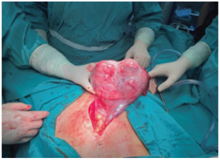
Figure 1: Intraoperative view of the tumor.
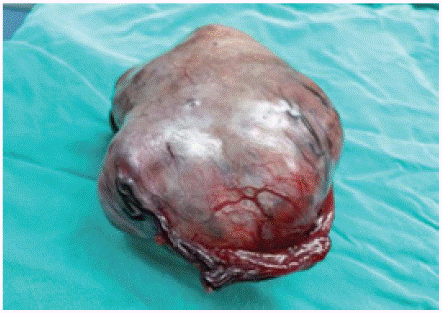
Figure 2: Gross appearance of huge pelvic tumor mass.
Postoperative pathohistological analysis has confirmed ovarian juvenile granulosa cell tumor with no tumor cell in the peritoneal washing. Omental and left ovarian biopsies have shown regular histomorphology. Cut surface of the tumor specimen showed compressed ovarian parenchyma by predominantly solid and partly cystic tumor tissue. Tumor cells were relatively monomorphic, round to oval and rarely spindle-shaped, with centrally placed vesicular nuclei and prominent nucleoli. One to two atypical mitoses were verified at 10 high power fields. Areas of cystic follicles were present, lined by single layer of flattened epithelium and lumens filled with basophilic content.
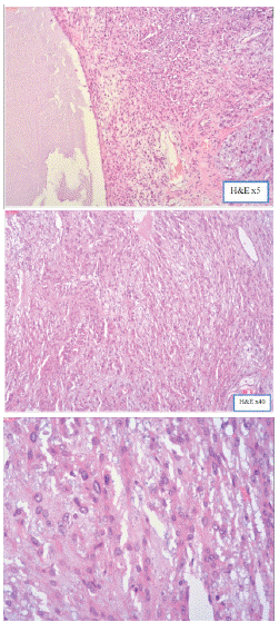
Figure 3a,b,c: Microscopic appearance of granulosa-cell tumor (Hematoxylin & Eosin stain).
Immunohistochemically, tumor cells showed positive signal for Calretinin, CD56, Inhibin, and negative signal for CD99 and WT-1. Ki-67 showed low proliferative index of 2-3% (Figure 4).
Figures 4a-4c. Immunohistochemical expression of granulolosa-cell tumor.
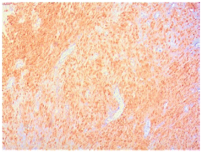
Figure 4a: Calretinin expression of tumor cells.

Figure 4b: CD56 expression of tumor cells.
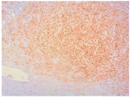
Figure 4c: Inhibin expression of tumor cells.
Considering the histological features, supported by the immunohistochemical findings, diagnosis of JGCT was made. To analyze the tumor stage, the International Federation of Gynecology and Obstetrics (FIGO) staging was used. On the basis of the FIGO classification, the tumor was categorized as stage IA.
Discussion
Juvenile granulosa cell tumorsare neoplasms which develop primarily in children and young adults, and approximately 90 percent are diagnosed before puberty.The mean age at diagnosis is 13 years of age, but patients’ age range from newborn stage to 67 years of age.In affected females, estrogen, progesterone, and testosterone levels may be elevated and lead to suppression of gonadotropins [3].
In our case there is no data for hormonal disorders. As a result, menstrual irregularities or amenorrhea are common, as in our case.
In addition to the hormonal changes, individuals may display tumor effects. Older patients usually seek medical attention for abdominal pain or swelling [3].
In our case there was abdominal distention which was the first symptom our patient has noticed.
Generally, 98% of JGCTs are unilateral. Solid consistency is predominant, but it may contain cystic component at various degree. In our case, the greatest part of the tumor was solid. The diameter of the tumor ranges from 2, 5 cm to 32 cm [4].
As JGCTs could reach big size, intraoperative rupture could be seen in 10% of cases [4]. Our patient presented with rapidly growing unilateral ovarian mass with diameter of 17 cm during the postpartal period and the unruptured tumor was excised totally during surgery.
These tumors usually grow slowly, but in this case the tumor rapidly reached big size. In our case, tumor had been diagnosed 18 months after delivery, thus the tumor presumably was present during pregnancy or even before. Most probably it has not been noticed during the pregnancy because of the initial small size which was masked by the growing uterus at regular ultrasound examinations. It may also be possible that the tumor appeared after pregnancy and has grown rapidly without any relation to our patients’ pregnancy. In general, JGCT is considered to have low malignant potential, but it has the risk of local extension and recurrences [5]. Almost all the recurrences of JGCT appear within 3 postoperative years [5]. The most important prognostic parameter is the stage of the disease [5]. Most of the cases are diagnosed at early stages (90% of cases at FIGO stage I) [4]. The five-year survival rate is about 95% in stage I and stage II tumors [5]. It is reported that excision of the tumor is almost always curative for stage IA tumors as it was in our case. However the course of the late stage tumors could be mortal [5,6].
In general, patients are young, and the involvement is unilateral, the removal of the involved ovary and uterine tube, and complete staging is the preferred surgical approach in order to preserve fertility [5]. Total abdominal hysterectomy and bilateral salpingo-oophorectomy are performed for late stage patients. Chemotherapy may be indicated [4]. After the surgery, the patient has been followed with regular gynecological examinations and until now there are no signs of regression.
Conclusion
JGCT may rapidly grow and reach big dimensions, and also could appear during the postpartum period. From all the data collected, we came to a conclusion that the tumor presumably was present during pregnancy or even before but had small dimensions and grew rapidly during the postpartum period, considering that during pregnancy despite all ultrasound data there was no evidence for the existence of the tumor. In our case, the tumor was categorized as stage IA and based to the regular gynecological examinations of the patient after the operation, until now there are no signs of regression, the prognosis appears to be excellent after surgical treatment with unilateral salpingo-oophorectomy to preserve fertility for our young patient.
References
- Liang Ma, Liwen Zhang, Yun Zhuang, Yanbo Ding, Jianping Chen. A rare case report of ovarian juvenile granulosa cell tumor with massive ascites as the first sign, and review of literature. Medicine (Baltimore). 2018; 97: e10916.
- Antovska V, Trajanova M. An original risk of ovarian malignancy index and its predictive value in evaluating the nature of ovarian tumour. SAJGO. 2015; 7: 52-59.
- Williams gynecology, 4e. 36: 771.
- Sahin S, Seçkin LSS. Postpartum Cystic Juvenile Granulosa Cell Tumor: A Case Report. 2014; 2: 30-36.
- Nucci MR, Oliva E. Gynecologic pathology. London: Elsevier Churchill Livingstone. 2009.
- Tavassoli FA, Devielee P. World Health Organization classification of tumours pathology and genetics of tumours of the breast and female genital organs. Lyon: IARC Press. 2003.