
Case Report
Austin Gynecol Case Rep. 2022; 7(2): 1032.
Enormous Pyosaplinx after Spontaneous Delivery
Antovska V1, Sozovska Belchovska E1, Vangelov A1, Antovich S2 and Dabeski D1
¹University Clinic for Gynecology and Obstetrics, Medical Faculty, St. Cyril and Methodius University Skopje, Republic of North Macedonia
²University Clinic for Abdominal Surgery, Medical Faculty, St. Cyril and Methodius University Skopje, Republic of North Macedonia
*Corresponding author: Dr. Eva Sozovska Belchovska, University Clinic for Gynecology and Obstetrics, Medical Faculty, St. Cyril and Methodius University Skopje, Republic of North Macedonia
Received: September 05, 2022; Accepted: October 10, 2022; Published: October 17, 2022
Abstract
Enormous pyosalpinx after a spontaneous childbirth is very rare event. Illness and progressive fever accompanied by abdominal or pelvic masses, vaginal purulent discharge, pain and pelvic discomfort, days to weeks after spontaneous delivery should add a suspicion for this condition. Ultrasound examination is the principle tool that can visualize enlarged tube.
In this article, we report a 38 year old woman with a post-partum enormous pyosalpinx in which the microbial agent was not identified. Progressively severe symptoms, such as vaginal discharge, abdominal and pelvic pain, malaise and high fever started 5 days postpartum.
An exploratory laparotomy showed left enormous pyosalpinx and massive adhesions with bowel and omentum. An excision of tubal ampoulae and omentum, as well as extensive lavage and drainage were made. Postoperative course was normal and the patient was dismissed in good condition.
Keywords: Pyosalpinx; Postpartal period; Puerperium
Abbreviations
Hgb: Hemoglobin; RBC: Red Blood Cells; WBC: White Blood Cells; Plt: Platelets; Hct: Hematocrit; Ne: Neutophils; Lymph: Lymphocites; Mono: Monocites; Gly: Glycose; Tot.Bil: Total Bilirubin; Dir/Ind Bol: Direct/Indirect Bilirubin; ALT: Alanine Aminotransferase; AST: Aspartate Aminotransferase; GGT: Gama Glutamil Tranferase; Alk: Phosp-Alkaline Phosphatase; S-A-amil: Serum Alpha Amylase; LDH: Lactate Dehydrogenase; CK: Creatin Kinase; K+: Potassium; Na++: Sodium; Fe: Iron; Alb: Albumins; Glob: Globulins; Tot.Prot: Total Proteins; CRP: C-Reactive Protein; PH: Power of Hydrogen; BE: Basal Excess; PT: Prothrombin Time; TT: Thrombine Time; aPTT: Activated Partial Thromboplastine Time; UA: Uric Acid; u (prefix): Urine
Introduction
Enormous pyosalpinx after vaginal spontaneous childbirth is very rare event. Illness and progressive fewer accompanied by abdominal or pelvic masses, vaginal purulent discharge, pain and pelvic discomfort, days to weeks after spontaneous delivery should add a suspicion for this condition. It is difficult to be sure whether the appendix or salpinx is inflamed. Ultrasound examination is the principle tool that could visualize an enlarged tube [1].
When PID follows pregnancy or an abortion, it is usually caused by a mixture of microorganisms, including anaerobes [2].
Case Presentation
In this article, we report a 38-year-old woman with postpartal enormous pyosalpinx in which no microbial agent was identified.
A 38-year-old woman was admitted to the University Gynecology and Obstetrics Clinic due to high fever, seven days after fourth vaginal delivery. She experienced a persistent fever (37.8-38.5°C) and liquid stools that were treated with probiotics, as well as left sided chest pain and dry cough. Her symptoms have deteriorated over time. Progressively severe symptoms started 5 days after delivery, at first with diarrhea and afterward with pleuropneumonia.
Her partner was healthy and also denied any history of sexually transmitted infections. At first, she has been admitted to the University Clinic for Invective diseases in Skopje, but after two days she was transferred to the University Clinic for gynecology and obstetrics.
On admission, the patient presented severely ill, with abdominal and pelvic pain, malaise and high fever (38.6°C), warm and pale skin with preserved hydration and elasticity, no efflorescence’s of infective genesis, firm right breast with warm and hyperemic skin, no palpable lymph nodes, dry lips, coated tongue, tonsils without patches, with normal pulse rhythm, eupnoic, followed by weakened medio-basal breathing. The palpation of the abdominal wall showed a suprapubic mass and tenderness in both iliac fossae. A pelvic examination revealed bulging cul-de-sac, and a tender pelvic mass. Chest X-ray showed consolidation of the left retrocardial parenchyma. There was a common side pleural effusion leading to diagnose of Pleuropneumonia.
The abdominal ultrasound and CT represented bigger amount of fluid in the left pleuro-costal sinus. The liver was normal in shape and size without any focal defects. There was a turbidity of the peripancreatic and mesenteric adipose tissue, more distally, near Culde- sac. The gallbladder, kidneys, adrenal glands and the bladder presented with normal shape, anatomy and function. The transvaginal ultrasound showed enlarged uterus with APD - 75mm, retroflected, with small residual mass in the cavity with diameter 9 mm. Next to uterus there was well delineated sausage-like formation partly solid, partly cystic, with dimensions 99x65 mm. There was small amount of free fluid in cul-de-sac.
The tumor marker Ca-125 showed slightly elevated levels of 77.2 UI/ml. The scoring system ROMI [3] showed low risk for ovarian carcinoma, e.g. ROMI =11 points: tumor size ≥6cm (1 point), cystic, with < ¼ solid parts (1 point), Opalescent intra-cystic fluid (2 points), septum ≥ 3 mm (1 point), vegetation of 3–5 mm (1 point), thickness of the capsule >5mm (2 points), serum levels of CA-125 in range of 35 – 129 U/ml (3 points). Doppler examination showed Resitence Index (RI) of 0,67, e.g. with low risk of the malignant nature of the tumor. The suspicion for benign pathology and big pyosalpinx was made. In the (Table 1) we represent the laboratory analysis.
![]()
Day of admission
First postoperative day
Fourth postoperative day
Hgb (g/L)
147
144
130
RBC (x10^12/L)
5.1
5.0
4.49
WBC (x10^9/L)
32.9
17.0
11.1
Plt (x10^9/L)
290
463
552
Hct (L/L)
0.41
0.42
0.36
Ne (%)
81
86
81
Lymph (%)
0.9
1
1.3
Mono (%)
0.5
0.4
0.5
Gly (mmol/L)
5.3
4.8
4.6
Urea (mmol/L)
10.1
3.4
1.1
Kreatinin (umol/L)
13.3
56
42
Tot. bil. (mmol/L)
7
10
7
Dir/Ind bil.(mmol/L)
2/5
3/7
2/5
ALT (U/L)
10
15
12
AST (U/L)
18
18
14
GGT (U/L)
27
39
26
Alk.Phosp. (U/L)
151
94
65
s-a-amil. (U/L)
52
221
180
LDH (U/L)
901
617
525
CK(U/L)
23
43
K+ (mmol/L)
3.0
3.5
3.5
Na++(mmol/L)
136
138
144
Fe(mmol/L)
1.2
3.8
3.4
Alb (g/L)
21
21
21
Glob
26
32
33
Tot. prot.
48
53
58
CRP (mg/L)
210
57
35
PH
7.39
7.44
PCO2
3.6
3.14
PO2
10.4
11.01
PHCO3
16.5
16.4
BE
-6.3
-5.3
O2 (%)
95.6
97.5
PT (sec.)
15
17
TT (sec.)
18
20
D-dimers
2718
>4500
u-PH
6.0
5.0
u-osm
1020
1020
u-prot
+
+
u-ket
+
-
Sediment
Renal epithel, bacteria, muscus
10-12 er
20-25 leBlood culture
Negative
Sputum culture
Candida albicans
Stool culture
Negative
C. difficile-negative
BAB (-)
HBsAg (-)
Anti-HCV (-)
Anti-HIV (-)
Table 1: Laboratory analysis of the patient with enormous postpartal pyosalpinx.
In (Figure 1) we represent the pelvic ultrasound on the admission.
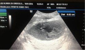
Figure 1: The pelvic ultrasound examination on the admission.
Immediately after admission, intravenous electrolyte solutions along with two antibiotics (Ceftriaxone 2g/daily and Metronidazole 1.5g in three daily doses), antipyretics, vitamins and anti-ulcer prophylaxis were administrated. Due to elevated platelets and d-dimer levels, thromboprofilaxis with enoxaparine was given.
Due to the patients’ condition and ultrasound findings, an urgent exploratory laparotomy was indicated. Surgery findings showed a firm tumorous formation connected by multiple adhesions with omentum and intestines. Partial adhesiolysis was performed in order to manage satisfactory access to the left ovary and fallopian tube, which were enlarged and filled with pus. The purulent content was drained and lavage with iodine was performed. Then the left fallopian tube and omentum were partially extirpated. The operative material was sent for histopathology analysis, and the pus was sent for microbiological analysis. Due to the presence of multiple adhesions, the inability to access the other pelvic organs and the distorted anatomy of the pelvis, further adhesiolysis was halted. Instead, one drain in the cavity of the fallopian tube was placed, and another one in the abdominal cavity. The surgery duration was approximately two hours, and estimated blood loss was 500mL.
The histopathology result was Salpingitis et peritonitis acuta fibrinopurulenta. The pus culture did not present with any pathogen bacteria, as was presumed since broad-spectrum antimicrobial treatment was previously administrated.
Postoperative treatment included intravenous antibiotic (Imipenem/Cilastatin), thromboprophylaxis with 40mg/day Enoxaparine, and fluid and blood components. The patient was discharged on the seventh postoperative day in good condition. She had been instructed to proceed with oral antibiotic (Cefixime) treatment for two more weeks. On the first six week follow-up, pelvic ultrasound showed absence of any pelvic tumor, left fallopian tube and left ovary were presented with normal shape and regular dimensions, as well as right adnexa and the uterus. In (Figure 2), we represent this early postoperative control ultrasound.
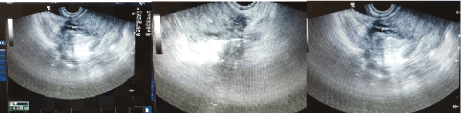
Figure 2: Six week follow-up pelvic ultrasound: A. Uterus and cul-de sac; B. Left adnexa; C. Right adnexa.
Discussion
According to Friedman and Bobrow [4], there are 4 mechanisms for infection of ovaries during pregnancy: haematogenous spread (pelvic tuberculosis), lymphatic spread, infection of a previously existing ovarian cyst and flare-up of old infection.
The preexisting salpingitis and flare-up of old infection, or lymphatic spread from vagina and cervix after delivery could be the mechanisms in our case. The adnexal infection shortly after vaginal delivery could be presented with some acute symptoms, like abdominal pain with giddiness and vomiting, which suggest peritonitis [5]. Since postpartal sexual intercourse with repercussive ascendant spread of infection in early postpartal period could be excluded, some systemic factors such as hematogenous or lymphatic bacterial spread, pelvic infection from adjacent organs or flare-up of old infection appeared to be taken into account as most probable mechanisms.
Pregnancy itself, especially the act of delivery when the internal uterine orificium is greatly dilated may be risk factors [5] which might promote favorable conditions for anaerobic invasion and growth in the uterus and adnexa. Many bacteria colonize lower genital organs, such as: Ureaplasma urealyticum, Mycoplasma hominus, group B Streptococcus (GBS), E. coli, Bacteroides spp, Gardnerella, Mobiluncus spp, and various enterococci, Chlamidia trachomatis, Neisseria gonorrhea, and could propagate upward and cause acute puerperal infection of the uterus and adnexa. They could stay asymptomatic for a long time and soar when the immune system of the host changes as in chronic diseases, trauma, starvation, pregnancy and especially after delivery. During pregnancy these organisms may enter the amniotic fluid either through intact choriodecidual membranes or after the membranes rupture and ultimately may infect the fetus and be a cause for a stillbirth [6]. Our patient has had 2 previous pregnacies with stillbirths.
The high virulence of the infective agent(s) and transient bacteremia, as well as decreased postpartal immunology status could be the cause of the systemic inflammatory response syndrome (tachycardia, tachypnea, dyspnea, high-grade fever, and high count of white blood cells), enterocolitis and pleuropneumonia, as well as present peritonitis which were present in our case [7]. The long-term high-dose preoperative antibiotic therapy could be the reason why the microbiology pus analysis came negative.
Conclusion
PID should be considered in a differential diagnosis of abdominal pain even during pregnancy, and especially in postpartal period, even though it is very rare. The preexisting salpingitis and flare-up of an old infection, or lymphatic spread from vagina and cervix after delivery could be the mechanisms of infection in this presented case. Prompt and early diagnosis is mandatory for decreasing maternal morbidity and mortality.
References
- William Silen, Zachary Cope - 2005 - Medical. Cope’s Early Diagnosis of the Acute Abdomen. https://books.google.com/books?isbn=019517545X
- Nelson Awori, Anne Bayley, Alan Beasley, James Boland, Michael Crawford, Frits Driessen, Allen Foster, Wendy Graham, Brian Hancock, Branwen Hancock, Gerald Hankins, Neville Harrison, Ian Kennedy, Julius Kyambi, Samiran Nundy, Joe Sheperd, John Stewart, Grace Warren, Michael Wood. Infection of the female genital tract; pelvic inflammatory disease [md] PID. The surgery of sepsis. Chapter 4. http://www.meb.uni-bonn.de/dtc/primsurg/ docbook/html/x1724.html
- Antovska V, Trajanova M. An original risk of ovarian malignancy index and its predictive value in evaluating the nature of ovarian tumour. SAJGO. 2015; 7: 52-59.
- Friedman S, Bobrow ML. Pelvic inflammatory disease in pregnancy. Obstetrics & Gynecology. 1959; 14: 417
- Navada HM, Bhat BP. Pelvic inflammatory disease in the form of peritoneal abscess complicating late pregnancy.Case Rep Obstet Gynecol. 2011; 2011: 851598.
- Goldenberg RL, McClure EM, Saleem S, Reddy UM. Infection-related stillbirths. Lancet (London, England). 2010; 375: 1482-1490.
- Duan R, Xu X, Wang X, Yu H. Perinatal outcome in women with bacterial sepsis: A cross-sectional study from West China. Medicine (Baltimore). 2019; 98: e17751.