
Case Report
Austin Gynecol Case Rep. 2025; 10(1): 1052.
Interstitial Ectopic Pregnancy: Case Study and Clinical-Surgical Approach: A Case Report
Chanaa I*, Azraq F, Bouchaib A and Alami MH
Les Orangers Maternity and Reproductive Health Hospital, Rabat, Morocco
*Corresponding author: Dr Imane Chanaa, Les Orangers Maternity and Reproductive Health Hospital, Rabat, Morocco Tel: +212 613 98 55 94; Email: Chanaa.gy@gmail.com
Received: May 12, 2025 Accepted: June 13, 2025 Published: June 16, 2025
Abstract
Background: To describe a rare form of ectopic pregnancy and the clinical, ultrasound, and surgical approach to this unusual form.
Methods: We report a case of interstitial pregnancy discovered by ultrasound in a 29-year-old multiparous woman with no particular history. The diagnosis was based on a clinical examination and an ultrasound performed by suprapubic and endovaginal routes. The therapeutic intervention was performed by laparotomy.
Results: The diagnosis of interstitial pregnancy was established using ultrasound data and confirmed by a βHCG level of 10,000 mIU/mL. On transparietal ultrasound, the gestational sac appeared intrauterine and fundal. On transvaginal ultrasound, the uterus was empty and enlarged, with a welldistinguished endometrial line. We identified an ectopic gestational sac, presented as a mass on the right side of the uterine fundus, measuring 21 mm in internal diameter, corresponding to approximately nine weeks of amenorrhea. This mass was surrounded by a 5 mm thick myometrium. No intraperitoneal fluid effusion was observed. The patient was managed by laparotomy, which was successfully performed.
Conclusions: Diagnosis of interstitial pregnancy is challenging, especially when relying solely on transparietal ultrasound, which is often insufficient to detect this pathology at an early stage. Prompt management is essential to prevent serious complications, such as uterine rupture. In the absence of immediate access to MRI in the emergency setting, transvaginal ultrasound, combined with βHCG measurement, constitutes a reliable method for early diagnosis and improved maternal prognosis.
Keywords: Ectopic pregnancy; HCG; Laparotomy
Introduction
Ectopic pregnancy (EP) corresponds to the implantation of the embryo outside the uterine cavity, generally in the fallopian tubes, but sometimes at the ovarian or abdominal level. Interstitial and ovarian locations, much rarer, represent specific diagnostic entities, with a frequency of 3.2% for ovarian pregnancies and 2.4% for interstitial [1]. It is characterized by the presence - on ultrasound, of myometrium around the gestational sac, cornual pregnancy can develop up to the 16th week, but exposes a high risk of sudden rupture and massive hemorrhage, making its prognosis more serious than that of classic tubal ectopic pregnancy [2]. Early and accurate diagnosis is therefore essential to prevent serious complications. Therefore, diagnosis is often based on a combination of clinical signs, plasma βHCG dosages and endovaginal ultrasound. Here we present the case of an unruptured interstitial pregnancy, detected early by endovaginal ultrasound in a 29-year-old patient.
Methods
Mrs. I. is a 29-year-old multiparous woman who has had two children delivered vaginally. The patient was not using hormonal contraception; moreover, the patient had used emergency contraception with levonorgestrel on several occasions. The reason for the consultation was metrorrhagia following nine weeks of amenorrhea. The history revealed right pelvic pain and sympathetic signs of pregnancy. The ultrasound examination was performed first by transparietal route and then by endovaginal route. On transparietal ultrasound, the gestational sac appeared intrauterine and fundal but not embryonated. The possible search for an embryo led us to use the endovaginal route. We observed by endovaginal route, a uterus of subnormal size, with a regular and decidualized cavity line, at a distance from a gestational sac in the form of a sessile ectopic fundal mass. This aspect was visible on longitudinal sections. On transverse sections, the gestational sac was in a right lateral position attached to the uterus, the cavity line of which was identifiable.
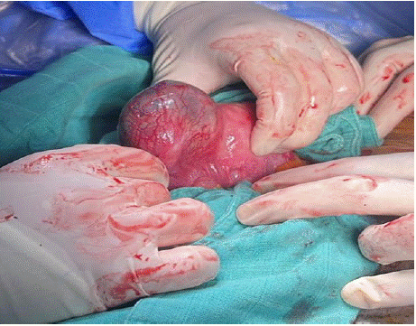
Figure 1: Interstitial Ectopic Pregnancy.
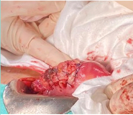
Figure 2: After laparotomy.
The ectopic gestational sac was non-embryonated and measured 21 mm in internal diameter . It was surrounded by a myometrium whose thickness measured 5 mm. There was no intraperitoneal fluid effusion. Both ovaries were individualized. The diagnosis of interstitial pregnancy was made on the basis of ultrasound data. It was confirmed by the measurement of plasma βHCG (10,000 mIU/mL) and the patient underwent a successful laparotomy. After aspiration of the hemoperitoneum estimated at 200 cc, we visualized the latero-uterine mass at the level of the right horn (Appendix I), after extraction of the pregnancy we performed the closure of the uterine cavity using 1-0 vicryl X-shaped stitches, hemostasis was ensured (Appendix II). The postoperative course was simple.
Results
The ectopic gestational sac was non-embryonated and measured 21 mm in internal diameter. It was surrounded by a myometrium whose thickness measured 5 mm. There was no intraperitoneal fluid effusion. Both ovaries were individualized. The diagnosis of interstitial pregnancy was made on the basis of ultrasound data. It was confirmed by the measurement of plasma βHCG (10,000 mIU/mL) and the patient underwent a successful laparotomy. After aspiration of the hemoperitoneum estimated at 200 cc, we visualized the latero-uterine mass at the level of the right horn (Appendix I), after extraction of the pregnancy we performed the closure of the uterine cavity using 1-0 vicryl X-shaped stitches, hemostasis was ensured (Appendix II). The postoperative course was simple.
Discussion
Clinical and Ultrasound Analysis
Interstitial pregnancy is an ectopic pregnancy of unusual location [3]. It is rare and represents less than 3% of ectopic pregnancy. Due to this low incidence, management is not codified. Several isolated cases have been reported in the literature [4]. Our case is a typical case of interstitial pregnancy diagnosed early. Our observation specifies the ectopic location of the mass attached to the uterine fundus. We agree with the data in the literature. According to these data, interstitial pregnancy gives an image of an abnormally eccentric ovular sac, surrounded by myometrium and protruding to the right or left of the uterine fundus [5,6]. Unlike isthmic pregnancy, where the ovular sac is separated from the uterine mucosa by the myometrium, the myometrium remains in direct contact with the mucosa in this case [7,8].
According to Timor-Tritsch [6], there are three essential criteria: 1- an empty uterine cavity, 2- a gestational sac separated by more than 1 cm from the uterine cavity (interstitial line), 3- a myometrial crown around this sac. We found these three criteria in our patient. These parameters proved to be relatively specific (88% to 93%), but their sensitivity was only 40% for the diagnosis of interstitial pregnancy [6]. They also spoke of the "interstitial line sign". This sign refers to the visualization of an echogenic line extending into the cornual region and reaching the middle part of the interstitial mass or gestational sac. In small interstitial pregnancies, this line may represent the interstitial portion of the tube. In larger interstitial pregnancies, it probably corresponds to the endometrial canal. The interstitial line sign is reported to have a sensitivity of 80% and a specificity of 98% for the diagnosis of interstitial pregnancy [6].
In our case, the diagnosis was not made by suprapubic pelvic ultrasound, which did not allow the pregnancy to be properly located. All authors agree that the endovaginal route is the best way to explore ectopic pregnancies [3,6,8]. According to Ackermann [7], endovaginal ultrasound is quite specific (88 to 93%) but its sensitivity, around 40%, is poor. Magnetic resonance imaging (MRI) is the most accurate alternative means for the positive and topographic diagnosis of rare forms of EP [5]. Classically, the βHCG level is often higher compared to tubal EP [9].
Treatment
Interstitial pregnancy has traditionally been treated by cornual resection or hysterectomy. This is probably due to the often-delayed diagnosis of this uncommon location. Thanks to advances in transvaginal ultrasound and more sensitive β-hCG tests, it is now possible to diagnose interstitial pregnancy at an earlier gestational stage, before rupture [10]. This opens new perspectives for conservative treatment by laparoscopy. Shapiro and Adler [11] were the first to report the successful excision of an ectopic pregnancy by laparoscopy. Today, laparoscopic treatment of tubal ectopic pregnancy has become the standard of care in hemodynamically stable patients. The first case of laparoscopic management of interstitial pregnancies was reported by Reich et al. in 1988 [12]. The techniques used included laparoscopic cornual resection with bipolar forceps and scissors, and ablation through colpotomic incision, as well as laparoscopic cornual resection with a unipolar knife after injection of vasopressin into the myometrium [12].
Acknowledgements
To all the authors who have contributed to the completion of this work.
Declarations
Funding: This work received no funding,
Conflict of interest: The authors declare no conflicts of interest,
Ethical approval: Informed consent has been obtained from the patient.
References
- Bouyer J, Coste J, Fernandez H, Pouly JL, Job-Spira N. Sites of ectopic pregnancy: a 10-year population-based study of 1800 cases. Hum Reprod. 2002; 17: 3224–3230.
- Jourdain O, Fontanges M, Schiano A, Rauch F, Gonnet JM. Prise en charge des autres ectopies annexielles (cornuale, inter- stitielle, angulaire, ovarienne). J Gynecol Obstet Biol Reprod. 2003; 32: 93–100.
- Ardaens Y, Guérin B, Perrot N, Legoeff F. Apport de l’échographie dans le diagnostic de la grossesse extra-utérine. J Gynecol Obst et Biol Reprod. 2003; 32: 3S28–13S.
- Bourdel N, Roman H, Gallot D, Lenglet Y, Dieu V, Juillard D, et al. Interstitial pregnancy. Ultrasonographic diagnosis and contribution of MRI. A case report. Gynecol Obstet Fertil. 2007; 35: 121–124.
- Poncelet E, Leconte C, Fréat-Martinez E, Laurent N, Lernout M, Bigot J, et al. Aspect échographique et IRM de la grossesse extra-utérine. Imag Femme. 2009; 19: 171–178.
- Timor-Tritsch IE, Monteagudo A, Matera C, Veit CR. Sonographic evolution of cornual pregnancies treated without surgery. Obstet Gynecol. 1992; 79: 1044–1049.
- Ackerman TE, Levi CS, Dashefsky SM. Interstitial line: sonographic finding in interstitial [cornual] ectopic pregnancy. Radiology. 1993; 189: 83-87.
- Fisch JD, Ortiz BH, Tazuke SI, Chitkara U, Giudice LC. Medical management of interstitial ectopic pregnancy: a case report and literature review. Hum Reprod. 1998; 13: 1981–1986.
- Wherry KL, Dubinsky TJ, Waitches GM, Richardson ML, Reed S. Lowresistance endometrial arterial flow in the exclusion of ectopic pregnancy revisited. J Ultrasound Med. 2001; 20: 335–342.
- Susie Lau MD and Togas Tulandi MD. Conservative medical and surgical management of interstitial ectopic pregnancy, FERTILITY AND STERILITYt. 1999; 72: 2.
- Shapiro HL, Adler DH. Excision of an ectopic pregnancy through the laparoscope. Am J Obstet Gynecol. 1973; 117: 290–291.
- Reich H, Johns DA, De Caprio J, McGlynn F, Reich E. Laparoscopic treatment of 109 consecutive ectopic pregnancies. J Reprod Med. 1988; 33: 885–890.