
Case Report
Austin Gynecol Case Rep. 2025; 10(1): 1051.
Migration of a Copper Intrauterine Device into the Sigmoid Colon: A Case Report
Chanaa I*, Azraq F, Bouchaib A and Alami MH
Les Orangers Maternity and Reproductive Health Hospital, Rabat, Morocco
*Corresponding author: Dr Imane Chanaa, Les Orangers Maternity and Reproductive Health Hospital, Rabat, Morocco Tel: +212 613 98 55 94; Email: Chanaa.gy@gmail.com
Received: May 12, 2025 Accepted: June 13, 2025 Published: June 16, 2025
Abstract
Background: We report the case of a 34-year-old patient, notion of insertion of an IUD two months ago in a health center, the evolution was marked by the installation of abdominal pain which motivated the consultation in our training for additional care.
Methods: We performed a first coelioscopy, the IUD was embedded in the sigmoid colon then we completed with a mini laparotomy for the removal and the revision of the sigmoid wound.
Results: We insist through this observation and in the light of the literature review on the efficacy and safety of the IUD when the technique and indications are rigorously respected, but also on one of the very rare complications of the insertion of the IUD.
Conclusions: The IUD is an effective contraceptive method; its insertion is a simple medical procedure that requires minimal knowledge and experience. Perforation is one of the rarest and most serious complications. Laparoscopy remains the most effective diagnostic and therapeutic method.
Keywords: IUD; Intraperitoneal migration; Coelioscopy
Introduction
Contraception by intrauterine device is one of the most used in the world, approximately 100 million users, it is a simple, effective and reversible method with a Pearl index of less than 1 per 100 woman years. Its contraceptive mode of action is located at the level of the tubes and spermatozoa, as well as at the level of the uterine cavity. However, their side effects, as well as their complications and contraindications, must be known to optimize its action [1, 2]. Perforation is one of the rarest and most serious complications, and which can cause the migration of the IUD in the various neighboring organs. Migrations have been described at the level of the cul-de-sac of Douglas, at the level of the omentum, the mesentery, and at the level of the bladder [2]. We report a new case of migration of the IUD in the sigmoid, the diagnosis of which was made 2 months after insertion. Ultrasound and unprepared abdomen were the means of diagnosis and the IUD, which was embedded in the sigmoid, was removed by mini-laparotomy.
Methods
Mrs. B, aged 34, is a multiparous woman, having 4 children delivered by VB, the last one dating back a year, having benefited from the insertion of a copper T-type intrauterine device two months ago. The patient did not undergo any follow-up until she consulted in our training for pelvic pain of the heaviness type following intense physical effort without any notion of bleeding or associated urinary or digestive signs. The clinical examination found the patient in good general condition, the abdomen was supple with slight tenderness on palpation. No visualization of the IUD strings on speculum examination. On vaginal examination, a normal-sized uterus was found to be slightly tender on palpation. The endovaginal pelvic ultrasound showed an empty uterus, we completed it with the abdomen without preparation (Figure 1) which highlighted the silhouette of the intrauterine device projected at the pelvic level, laterally deviated to the left, absence of pneumoperitoneum
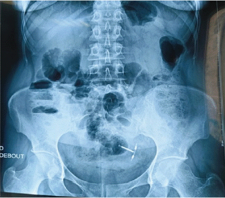
Figure 1: The ASP in standing position.
Results
The diagnosis of secondary perforation of the uterus and partial migration of the IUD was retained and the patient underwent a laparoscopy for removal of the migrating IUD. The exploration allowed visualization of the arms of the IUD through the sigmoid colon with an omental inflammatory reaction. After discussion with the visceral surgeon, a mini-laparotomy of the Pfannenstiel type was decided, thus allowing visualization of the IUD whose vertical branch was implanted in the intestinal lumen. (Figure 2). The IUD was removed and then the sigmoid defect was closed with several interrupted stitches and a running suture. The postoperative course was unremarkable.
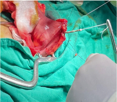
Figure 2: Image of the IUD embedded in the lumen of the sigmoid colon.
Discussion
The intrauterine device is the most widely used method of contraception in the world, with approximately 100 million users. It is a contraceptive method that uses a mechanical process of local action. There are currently several types of IUDs, the inert IUDs (Lippes Loop) which are no longer used, and the bioactive IUDs, made of copper, copper-silver or progestins are the most widely used due to their better tolerance [1].
IUD insertion is a simple medical procedure, governed by legal obligations and laws, requires a minimum of medical knowledge and a minimum of practice because in certain situations, this insertion can be followed by complications such as infection or uterine perforation [1, 2]. The incidence of perforation is rare, it does not exceed 1.3 per 1000 insertions, according to large clinical trials reported [3–5]. These perforations can be partial, when only part of the IUD pierces the uterine wall or the cervix, or complete, when the IUD crosses the uterine wall to enter the abdominal cavity [2–5]. It most often occurs at the time of insertion, but it can go unnoticed and be discovered only secondarily [6].
Several factors can intervene and be at the origin of the perforation, first of all uterine factors, with a small size, a significant malposition, in particular a retroversion, a fragility of the myometrium by multiple pregnancies, hypoplastic uteri, scarred uteri, factors linked to the insertion in particular which requires a push, and the inexperience or clumsiness of the operator [1, 2]. On the physio-pathological level the importance of the endometrial inflammation caused by this foreign body prevents implantation.
This inflammation is a double-edged sword, causing a significant accumulation of enzymes and lysosomal lytic substances promoting endometrial destruction and IUD migration [7] as is the case of our patient where a significant inflammatory reaction was highlighted. After perforation, the device can, and in the majority of cases, remain in the pelvic cavity, migrate into hollow organs, particularly in the bladder, or be surrounded by the omentum and remain inert for several years, especially for non-active IUDs [8, 9]. In the literature we have counted more than 120 cases of intra-abdominal migration of the IUD, 59 cases of intra-vesical migration; extra-vesical and abdominal pelvic migrations as in the case of our patient are exceptional [2–10]
Clinically, the symptomatology varies depending on the location of migration and the type of IUD. Uterine perforation by an IUD is usually asymptomatic. Except when it is concomitant or just after insertion, as was the case in our patient who experienced an exacerbation of pain two months after insertion.
An unprepared abdominal X-ray confirms expulsion if the IUD is not found on the image. Its visualization does not prejudge its situation [9]. In our case, the ASP confirmed the existence of the device, which was laterally deviated to the left. Suprapubic ultrasound is a fundamental step in the diagnosis. It allows visualization of the IUD intrauterine or in another location [2–9]. In cases where the paraclinical diagnosis is difficult, a laparoscopy or diagnostic laparotomy may be of crucial importance.
At a distance from the insertion, the indication for the removal of an IUD is imperative and must be carried out quickly due to the above-mentioned complications which increase the risk of female morbidity and mortality [9]. The treatment of intra-abdominal migrations remains removal by coelioscopy or laparotomy [9, 10]. In our case, a coelioscopy was performed and which revealed a copper IUD embedded in the sigmoid and which was removed without incident.
Acknowledgements
To all the authors who have contributed to the completion of this work.
Declarations
Funding: This work received no funding,
Conflict of interest: The authors declare no conflicts of interest,
Ethical approval: Informed consent has been obtained from the patient.
References
- Boudineau M, Multon O, Lopes P. Contraception par dispositif intra-utérin. Encycl Méd Chir- Gynécologie. 2001; 738-A-09: 7.
- Zouhal A, el Amrani N, Bensaid F, et al. Migration intra-vesicale d'un dispositif intra-uterin a propos d'un cas. Rabat, Maroc: Maternité Universitaire des Orangers; 2000-2001.
- Treiman K, Laurie Liskin SCM, Adrienne Kols, et al. Les DIU: état récent des informations. Population Reports (Series B). 1995; 6: 1–35.
- Ledward RS, Healey C, Eadie R. Removal of extrauterine Saf-T-Coil through laparoscope. British Medical Journal. 1972; 1: 508.
- Zakin D, Stern WZ, Rosenblatt R. Complete and partial uterine perforation and embedding following insertion of intrauterine devices I Classification, complications, mechanism, incidence, and missing string. Obstetrical & Gynecological Survey. 1981; 36: 335–353.
- Gruber A, Rabinerson D, Kaplan B, Pardo J, Neri A. The missing forgotten intrauterine contraception device. Contraception. 1996; 54: 117–119.
- Chang CH, Chou CY, Lee WI, Tzeng CC, Liuc H. Pelvic actinomycosis with colo-ileo-vesical fistula formation: report of case. J Formos Med Assoc. 1992; 91: 342–345.
- Markovitch O, Klein Z, Gidoni Y, Holzinger M, Beyth Y. Extrauterine mislocated IUD: is surgical removal mandatory. Contraception. 2002; 66: 105–108.
- Brar R, Doddi S, Ramasamy A, Sinha P. A forgotten migrated intrauterine contraceptive device is not always innocent: a case report. Case Rep Med. 2010: pii–740642.
- Bacha Khaled, Ben Amna Marouane, Ben Hassine Lofti, et al. Dispositif intrautérin migré dans la vessie. Progrčs en Urologie. 2001 ; 11: 1289–1291.