Case Report
Austin Gynecol Case Rep. 2025; 10(1): 1050.
Antenatal Diagnosis of Thanatophoric Dwarfism: About a Case Report and Review of the Literature
Chanaa I*, Houjjaj D, Bouchaib A, Alami MH
Les Orangers Maternity and Reproductive Health Hospital, Rabat, Morocco
*Corresponding author: Dr Imane Chanaa, Les Orangers Maternity and Reproductive Health Hospital, Rabat, Morocco Tel: +212 613 98 55 94; Email: Chanaa.gy@gmail.com
Received: May 12, 2025 Accepted: June 13, 2025 Published: June 16, 2025
Abstract
Thanatophoric dwarfism is a rare osteochondrodysplasic classified into two types I and II. It is due to a mutation in the FGFR3 (fibroblast growth factor receptor 3) gene located on the short arm of chromosome 4. This morphological anomaly is always lethal, and molecular biology is used to diagnose it with certainty. We report the case of a 25-year-old nulliparous woman with no particular history, whose ultrasound scan at 38 weeks’ amenorrhea, performed as part of standard prenatal surveillance, led to the diagnosis of NT type I in the face of highly suggestive fetal dysmorphic images. These included macrocephaly and extremely shortened limbs associated with curved femurs. A 33 cm long, 2600 g dwarf neonate was extracted from the pelvis and admitted to the neonatal intensive care unit for severe respiratory distress at birth, with death at 7 days.
Background: Thanatophoric dwarfism is a rare osteochondrodysplasic classified into two types I and II. It is due to a mutation in the FGFR3 (fibroblast growth factor receptor 3) gene located on the short arm of chromosome 4.
Methods: We report the case of a 25-year-old nulliparous woman with no particular history, whose ultrasound scan at 38 weeks’ amenorrhea.
Results: A 33 cm long, 2600 g dwarf neonate was extracted from the pelvis and admitted to the neonatal intensive care unit for severe respiratory distress at birth, with death at 7 days.
Conclusions: NT is a major fetal morphological anomaly for which antenatal diagnosis is imperative. In the absence of molecular biology, obstetrical ultrasonography, sometimes coupled with radiography of the uterine contents, enables NT to be diagnosed and other types of micromelic dwarfism to be ruled out.
Keywords: Thanatophoric dwarfism; Morphological anomaly; Surgery; Case report
Introduction
Thanatophoric dwarfism (NT) is a rare osteochondrodysplasic classified into two types I and II. It is due to a mutation in the FGFR3 (fibroblast growth factor receptor 3) gene located on the short arm of chromosome 4. This morphological anomaly is always lethal, and molecular biology is used to diagnose it with certainty. But medical imaging must be at the forefront of early prenatal screening. We report the case of a 25-year-old nulliparous woman with no particular history, whose ultrasound scan at 38 weeks' amenorrhea, performed as part of standard prenatal surveillance, led to the diagnosis of NT type II in the face of highly suggestive fetal dysmorphic images. Highroute delivery for pelvis limitation enabled extraction of a dwarf neonate measuring 33 cm in length and 2600 g in weight, who was hospitalized in the neonatal intensive care unit for severe respiratory distress at birth.
Methods
Patient information: Mrs HM, aged 25, G1P1, was admitted to the Ibn Sina Hospital in ORANGERS for a prognosis of 38SA delivery. There was no known family history of congenital malformation, nor any notion of constitutional short stature in the couple or consanguinity. The patient underwent ultrasonography at 15SA, which revealed no fetal skeletal malformations.

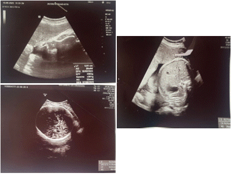
    Figure 1: obstetrical ultrasound at 38 weeks’ gestation showing: a curved
femur (A) with macrocephaly (B), with abdominal macrosomia (C).

Figure 2: Showing a characteristic aspect of thanatophoric dysplasia.
Results
Diagnostic approach: ultrasonography at 38 SA revealed a female fetus with a BIP of 102.5 mm greater than 90 of gestational age, a poorly developed thorax an abdomen of normal volume (DAT=107.7, CA=360.1 mm greater than 90 of gestational age) and, above all, shortening of the bones of all four limbs with femurs that are curved: LF= 50.1 mm below the 10 percentile of gestational age and with an LF/PIED ratio <0.8, note the presence of hydramnios with a large cistern at 100mm. There were no other associated morphological abnormalities. The diagnosis of type II TD was made on the basis of these findings, and the couple was informed.
Therapeutic intervention and follow-up: delivery was via a high route for a borderline pelvis and unfavorable bi-spinous BIP confrontation, giving birth to a female newborn, PDN= 2600g ap 10, who presented respiratory distress for which she was hospitalized in the neonatal intensive care unit.
Discussion
NT is a major and lethal fetal malformation. According to Machado et al [1], the term thanatophore is derived from the Greek myth "thanatos" and means personified death. The life expectancy of newborns with NT is estimated at around one hour after birth by Noe et al. [2], who have evoked some rare cases of survival up to five and eight years. In our observation, the newborn with NT presented respiratory distress. This will be related to the narrowness of the rib cage with a lack of lung development would be involved according to Pietryga et al. [3].
Our patient was 25 years old with no particular history. The notion of consanguinity or related couple noted in the literature was not found in our case. In the study by Lahmar-Boufaroua in Tunisia, consanguinity was found in 61% of cases.
Antenatal diagnosis of fetal malformations whether lethal or not should be the haunt of every sonographer. To this end, some authors recommend early morphological anomaly detection during firsttrimester ultrasound [4]. This ultrasound should insist on nuchal clarté. According to Tonni et al [5], nuchal hyperclarity is an early sign of NT. In our observation, the sonographer had only measured the thickness of the nuchal translucency without evoking its echogenicity.
We believe that the quantity of amniotic fluid should also be taken into account in ultrasound exploration during the prenatal check-up. Indeed, hydramnios is a clinical and ultrasound sign consistently found in the literature as part of the signs associated with NT [5]. We observed it, in our case.
Signs of NT and classification as type I or II are evident from the second trimester onwards on two-dimensional ultrasound. These include macrocrania, a narrow thorax, a prominent abdomen and extremely short limbs. In type I, the femur is curved, whereas in type II it is not. Recent advances in technology, with the advent of threedimensional ultrasound, have further facilitated the diagnosis of NT [6]. However, the fear of making a mistake or confusing NT with achondroplasia, osteogenesis imperfecta, achondrogenesis or any other non-lethal micromelia means that ultrasound must be coupled with molecular biology, which provides a diagnosis of certainty [7]. In its absence, it would be advisable to combine ultrasound with an X-ray of the uterine contents [8]. Noe et al [9] estimated vertebral compression and shortening at around one hour after birth, and reported rare cases of survival up to five and eight years. According to Pietryga et al.[10], severe respiratory distress linked to the narrowness of the thoracic cage and lack of lung development is the cause.
Acknowledgements
To all the authors who have contributed to the completion of this work.
Declarations
Funding: This work received no funding,
Conflict of interest: The authors declare no conflicts of interest,
Ethical approval: Informed consent has been obtained from the patient.
References
- Wilcox WR, Tavormina PL, Krakow D, Lachman RS, Wasmuth JJ, Thompson LM, et al. Molecular radiologic and histopathologic correlations in thanatophoric dysplasia. Am J Med Genet. 1998; 78: 274-281.
- Kocherla K, Kocherla V. Antenatal diagnosis of Thanatophoric Dysplasia: a case report and review of literature. Int J Res Med Sci. 2014; 2: 1176-1179.
- Lahmar-Boufaroua A, Yacoubi MT, Hmisssa S, Selmi M, Korbi S. Les ostéochondro dysplasies létales: étudefoeto pathologique de 32 cas. Tunisie Med. 2009; 87: 127-132.
- Noe EJ, Yoo HW, Kim KN, Lee SY. A case of thanatophoric dysplasia type I with an R248C mutation in the FGFR3 gene. Korean J Pediatr. 2010; 53: 1022-1025.
- Tirumalasetti N. Case report of Thanatophoric dysplasia: A lethal skeletal dysplasia. J NTR Univ Health Sci. 2013; 2: 275-277.
- Brodie SG, Kitoh H, Lipson M, et al. Thanatophoric dysplasia type I with syndactyly. Am J Med Genet. 1998; 80: 260-262.
- Petitcolas J, Couvreur A, Leboullenger P, Rossi A. Intérêt de l’échographie morphologique précoce pour la détection des anomalies chromosomiques. J Gynecol Obstet Biol Reprod. 1994; 23: 57-63.
- Tonni G, Azzoni D, Ventura A, Ferrari B, Felice CD, Baldi M. Thanatophoric dysplasia type I associated with increased nuchal translucency in the first trimester: early prenatal diagnosis using combined ultrasonography and molecular biology. Fetal Pediatr Pathol. 2010; 29: 314-322.
- Phatak SV, Pandit MP, Phatak MS, Kashikar R. Antenatal sonography diagnosis of thanatophoric dysplasia: A case report. Indian Journal of Radiology and Imaging. 2004; 14: 161-163.
- Lingappa HA, Karra S, Aditya A, Batra N, Chamarthy NP, Ravi Chander KWD. Autopsy Diagnosis of Thanatophoric Dysplasia. J Indian Acad Forensic Med. 2013; 35: 296-298.