Department of Medicine, Rutgers University, USA
*Corresponding author:Dharmesh H Kaswala, University Hospital, Rutgers University, New Jersey Medical School, Department of Medicine, Division of Gastroenterology and Hepatology, Newark, NJ, USA
Received: October 20, 2014; Accepted: October 22, 2014; Published: October 24, 2014
Citation: Kaswala DH and Ahlawat S. Traumatic Hemobilia - An Image Challenge. Austin J Gastroenterol. 2014;1(5): 1025. ISSN:2381-9219
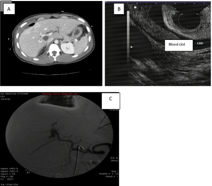
A 24 year old man admitted to hospital status post traumatic knife injury causing traumatic hemopneumthorax and liver laceration. Patient underwent bilateral chest tube placement and operative repair of liver laceration involving right lobe of the liver. After 2 weeks of the hospital stay patient found to have abnormal liver function tests; AST: 130, ALT: 86, T.Bil:1.2, Alk phos: 1128, Albumin: 2.2, Direct Bili: 0.7. Hepatitis serology for B and C was negative. Patient’s CT scan of the abdomen, EUS, ERCP and cholangiogram images shown. Hepatic angiogram showed on Image C.
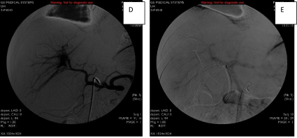
Traumatic hemobilia consists of hemorrhage into the biliary tract as a result of abdominal trauma. The classical triad of biliary colic, jaundice and upper gastrointestinal bleeding is not a constant finding, and clinically silent hemobilia has been reported. Accidental liver injury is the second commonest cause of hemobilia, exceeded only by iatrogenic causes. However, hemobilia is an uncommon complication of trauma and occurs in less than 3% of all hepatic injuries [1,2]. Hepatic parenchymal injuries, such as a liver laceration and hematoma, are often accompanied by traumatic hemobilia. Blunt injury is the most common mechanism in post-traumatic hemobilia [3]. The cause of bleeding found to be heaptic artery to portal vein fistula (Image D) leading to traumatic hemobilia. By Interventional Radiologist the right hepatic artery portal venous fistula emboli zeds with 5mmx5mm coil (Image E). The follow up imaging study demonstrated right hepatic artery occluded. The fistula to the portal vein is no longer enhancing. The left hepatic artery was widely patent. The delayed imaging into the portal venous phase showed the portal vein is patent. After intervention the liver enzymes went back to normal in few days. In this case the cause of obstructive jaundice was CBD thrombosis due to traumatic hemobilia.
In our case there was a fistula between hepatic artery and portal vein due to traumatic knife injury which must have caused injury to bile duct causing blood clot in CBD but the site of fistula between portal vessels and biliary system may have been sealed off. Although ultrasonographic study is usually normal in patients with hemobilia, it can detect intrahepatic hematomas and blood clots in the gallbladder, and is also helpful in evaluation of other solid organs. Abdominal CT scan is a useful modality in cases of abdominal trauma, and may be helpful in recognition of accompanying parenchymal disruptions. However, early-stage traumatic hemobilia may not be detected by CT scan because the amount of bleeding is too small. Endoscopy and ERCP can also be used to diagnose traumatic hemobilia. Traumatic hemobilia should be managed by vascular embolization, surgery, or endoscopic management because hemobilia may rarely disappear spontaneously. Endoscopic therapy had shown as a safer alternative to existing methods [4].
Image A: CT scan showing blood clot; Image B: Endoscopic Ultrasound with blood clot and Image C: Hepatic angiogram.

Fluroscopic view of Rt hepatic artery and portal venous fistula:
Image D: Pre embolized ; Image E: Post embolized.
