
Introduction
Parvovirus B19 (PVB19) is one of the smallest viruses that are known to infect humans [1]. The clinical manifestations of B19V infection vary greatly and depend on age, hematologic and immunologic status. In immunocompetent individuals, the infection can be completely asymptomatic or can cause mild and self-limiting clinical manifestations such as erythema infectiosum or fifth disease during childhood, arthralgias and arthritis in adults, particularly in women [2,3], chronic hemolytic anemia, and fetal death in utero or non-immune hydrops fetalis in pregnant women [4]. Due to the efficient replication of B19V in the erythroid progenitor cells [5].
In temperate climates, the infection may occur throughout the year, Infection is most common in late winter or early spring [6]. During pregnancy, the virus is transmitted through exposure to infected respiratory droplets or blood products and vertically from mother to fetus [7,8]. The vertical transmission of B19 occurs in about one-third of women infected [9].The proportion of pregnant women susceptible to B19 infection ranges from 34% to 65% in various parts of the world. The incidence of seroconversion during pregnancy is estimated at between 1% and 1, 5% in the endemic period, increasing to 13% in the epidemic period [10]. Non-immune women are most likely to be infected by young children [11].
Parvovirus infection of mothers is diagnosed using serologic or an immune assay enzyme B19 IgM and B19 IgG [12]. Viral DNA can be detected by Polymerase Chain Reaction (PCR) and is considered to be the best indicator of infection in maternal, fetal blood, and amniotic fluid [13].
In these conditions, it seemed necessary to us to study this virus in this article and to answer the various practical questions raised by the occurrence of contagion and/or infection with Parvovirus B19 during pregnancy.
Parvovirus B19: A Few Words of History
Parvovirus B19 particles were first described in 1975 by Cossart, an Australian virologist working in London [1]. While checking normal blood donor’s serum in an assay for hepatitis B she noticed an anomalous reaction in position 19 plate B [14]. This virus was successively called SPLV (Serum Parvovirus like Virus), Aurilac antigen (in France), Nakatani antigen (in Japan), then B19, number of the blood bag where it was isolated for the first time. This explains the name of the virus when there is no parvovirus B1 to B18.The pathogenic role of the virus was first identified in 1981 during erythroblastopenia attacks in patients with sickle cell disease. In 1983, it was recognized as the cause of erythema infectiosum or the 5th pediatric disease [15]. Subsequently, PVB19 infections have been linked to fetoplacental hydrops, Fetal Deaths in Utero (MFIU) secondary to fetal anemia or myocardial involvement [16].
Virology and Pathophysiology of B19V Infection
B19 virus: Virology
Taxonomy: Parvoviruses are common animal and insect pathogens. The Parvoviridae family is divided into two sub-groups:the Parvovirinae infecting vertebrate cells, and the Densoviridaeinfecting invertebrate cells (Table 1) [17]. The Parvovirinae are further subdivided into three groups:
![]()
    Genus
    Virus
    Natural hosts(s)
    Clinical spectrum


    Parvovirus
    Aleutian    mink disease virus
    Mink, ferret, skunk,raccoon
    Immune complex disease and fetal death


    Canine    parvovirus
    Dog
    Enteritis, myocarditis


    Mice    minute virus
    Mouse, rat
    No known disease


    Porcine    parvovirus
    Pig
    Abortion, fetal death


    Dependovirus
    Adeno-associated    virus 1 to 6
    Human
    No known disease


    Avian    adeno-associated virus
    Birds
    No known disease


    Canine    adeno-associated virus
    Dog
    No known disease


    Bovine    adeno-associated virus
    Cow
    No known disease


    Erythrovirus
    Parvovirus    B19
    Human
    Erythema infectiosum, aplastic crisis, arthritis,    hydrops fetalis, etc.


    Parvovirus    V9a
    Human
    Aplastic crisis?


    Chipmunk    parvovirusa
    Chipmunk
    No known disease


    Simian    Parvovirusa
    Cynomolgus monkeys
    Anemia


    Pig-tailed    macaque Parvovirusa
    Pig-tailed macaques
    Anemia and immunosuppression


    Rhesus    Parvovirusa
    Rhesus monkeys
    Anemia


    a Proposed member of genus.
Table 1: Excerpt of the current classification of the subfamily Parvoviridae, including proposed members of the genus Erythrovirus placed tentatively [17].
• Genus Parvovirus that replicate autonomously.
• Genus Dependovirus that needs helper viruses to replicate.
• Genus Erythrovirus that need erythroid cells to replicate.
Parvovirus B19 belongs to the genus Erythrovirus.
Morphology: Parvovirus B19 is a small single-stranded DNA virus [18]. Parvovirus B19 is a non-enveloped, 22 to 26 nm icosahedral virus (Figure 1), containing a single strand of DNA of approximately 5,500 nucleotides. As with other parvoviruses, B19 employs overlapping reading frames to encode on-structural proteins and two capsid proteins. The B19 virion is an icosahedron consisting of 60 copies of the capsid proteins.
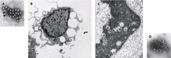
    Figure 1: Transmission Electron Micrographs Showing Native Parvovirus B19 in Serum and Cells and Recombinant Capsid [14].
Panel A shows symmetric, icosahedral particles, about 25 nm in diameter, and empty capsids (arrowheads) in serum from an infected. Panel B shows human
erythroid progenitor cells infected in vitro with the virus; vacuoles and cytoplasmic pseudopods are present (×10,000). In Panel C which shows part of Panel B
at higher magnification, marginated chromatin contains assembled capsids (×100,000). Panel D shows empty recombinant parvovirus capsids produced in a
baculovirus system (×154,000).
Most of the capsid consists of VP2, the major structural protein (molecular weight 58 kDa), with 5% or less of the larger VP1 protein, the minor protein (83kDa). Using genetic engineering techniques, the capsid proteins have been expressed in a variety of both mammalian and insect cell lines, where they self-assemble in the absence of DNA and form recombinant empty capsids [19]. VP1 is not required for capsid formation. The limited DNA content and the absence of a lipid envelope makes this virus resistant to heat (56oC for 60 min) and lipid solvents [20].
Genomic structure and organization: As in most animal Parvoviruses, the B19 genome has two large open reading frames, with the single Nonstructural Protein (NS1) encoded by genes on the left side of the genome and the two capsid proteins (VP1 and VP2) by genes on the right side. The non-structural protein, from NS1, sub serves multiple replicative functions and is cytotoxic to host cells [21]. The two structural proteins, Viral Protein1 (VP1) and Viral Protein 2 (VP2), arise from alternative splicing so that VP1 is the same as VP2 except for an additional 226 amino acids at its amino-terminal (Figure 2) [22-24].
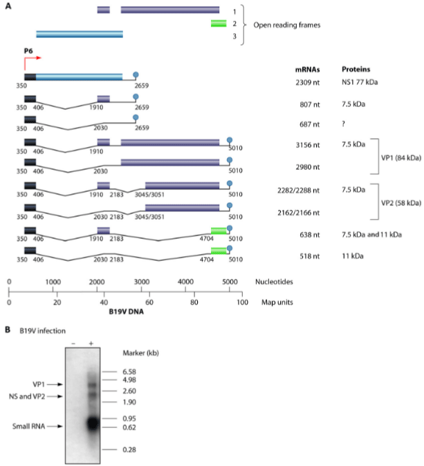
    Figure 2: Schematic Representation of B19V Genome Organization and Functional Mapping [22-24].
(A) Transcription map of B19V genes. The 9 main transcripts are represented. (B) Expression of viral mRNAs in primary erythroid cells. The viral mRNAs were
detected by Northern blotting, using a radiolabeled probe in non-infected and infected primary erythroid cells as described previously.
NS1 is a 671 amino acid long protein that has an MW of ~75 kDa. NS1 contains two nuclear localization signals. NS1 contains a DNA binding and endonuclease domain at the N-terminus
In short, NS1 is a multifunctional protein and plays various roles during B19V infection [25]. VP2 is the predominant protein, comprising 95% of the virus capsid. VP1 is the same as VP2 except for an additional 226 amino acids at its amino-terminal [26]. VP1makes up only 5% of the capsid, has its unique region external to the viral capsid itself, and is thought to be the main target of neutralizing antibodies. Sequence analysis reveals that NS1 is highly conserved, while VP1and VP2 show greater variation [27]. Despite variations in VP1 and VP2, the antigens are commonly and successfully used in serologic tests.
B19V as a species is subdivided into three genotypes, the prototype genotype 1, and two variant genotypes 2 and 3. At the nucleotide level, the diversity between genotype clusters is about 10%, while the diversity within each genotype cluster is normally lower than 2% for genotype 1 and in the range 3-10% for genotypes 2 and 3 [28]. All genotypes co-circulate, but with different frequencies and geographical distributions [29].
Pathogenesis and Infection
Viral life cycle
The only known natural host cell of parvovirus B19 is the human erythroid progenitor. Like other non-enveloped DNA viruses the Parvovirus B19 life cycle includes the following stages: binding to host cell receptor, internalization, translocation of the genome to the host nucleus, DNA replication, RNA transcription, assembly of capsid, packing the genome, and cell lines is with release of the mature virion [3] (Figure 3). The P antigen on the red blood cell is a cellular receptor of the Parvovirus B19.However, all P antigen expressing cells are not permissive to B19V. Various other co-receptors like integrina5β1, and antibody-mediated B19V entry routes are presumed to be involved in B19V entry [30].
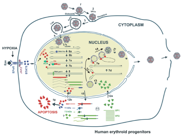
Figure 3: Proposed model of B19V life cycle [31].
Pathogenesis and Immune Response
After primary infection, mild symptoms of fever and general illness usually start after 6-10 days when viremia is highest. These symptoms will recede within a week [32,33]. During the second week after infection, the viremia titer decreases, and IgM antibodies are detected. In the third week, the well-known slapped cheeks and rash and the possible arthralgia occur, which coincides with an IgG antibody response [34]. These IgG antibodies can persist for years, while the IgM antibodies disappear after 6-10 weeks (Figure 4).
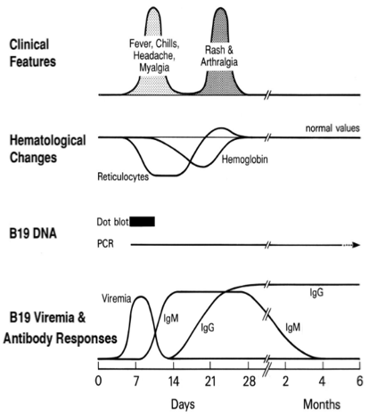
Figure 4: Virologic, immunologic, and clinical course following B19 infection [37].
Recovery involves production of IgM antibody 10 to 12 days post infection, coinciding with a peak in virus level. IgM usually persists in serum samples for approximately 3 months but may be found for several months [35]. IgG antibody is detectable in volunteers about 2 weeks after inoculation and presumably persists for life and protects against secondary infections. IgA may also be detected and probably plays a role in protection against infection by the natural nasopharyngeal route [36].
Parvovirus B19 and Autoimmunity
Apart from Rheumatoid Arthritis (RA), B19 infection has been associated with the onset of numerous autoimmune disorders including Systemic Lupus Erythematosus (SLE) [38], other connective tissue diseases, and systemic vasculitides. Although a few cases of erosive RA and SLE have been associated with B19 infection, the virus is probably an extremely rare cause of these diseases. Systemic vasculitides including, for example, Henoch-Schönlein purpura, periarteritis nodosa, and giant cell arteritis can occur after acute B19 infection [39]. The role of B19 in these disorders is not clear and in some cases, the infection may be a pure coincidence and in other cases, it can be a triggering or even a rare aetiological factor [40].
Parvovirus B19 Infection in Pregnancy
Acute parvovirus B19 infection is a risk for pregnant women. Since B19 infection occurs mainly during childhood, children represent a main source for virus transmission.
Human parvovirus B19 infection is widespread. Approximately 30-50% of pregnant women are nonimmune, and vertical transmission is common following maternal infection in pregnancy. The magnitude of B19 has been studied in many developed countries [41] whereby the prevalence of specific B19 antibodies among pregnant women has been found to range from 1 to 5% with a transmission rate to the fetus of about 17-33% [42].
Approximately 50% to 75% of women of reproductive age have developed immunity to parvovirus B19 [43,44]. Without known exposure, about 1% to 3% of susceptible pregnant women will develop serologic evidence of infection in pregnancy [45]. Rising to over 10% in epidemic periods [46]. Where there is extensive opportunity for exposure to parvovirus B19, such as in a daycare center or school, it is estimated that 20% to 30% of susceptible women will develop an infection, while 50% of susceptible women exposed through household contacts will become infected [47].The risk of infection in pregnant women with one child is (are)3 times more than nulliparous women, but this risk for women with three or more children is (are) 7,5 times more. The other risk factors are working in the school, care centers, and other full stress jobs [48,49].
Fetal Effects of Parvovirus B19 Infection
Primary PVB19 infection during pregnancy is potentially fatal to the fetus. Transplacental passage would occur at the time of the maximum peak of viremia following maternal contamination approximately one week. It is made possible by the expression of the P antigen on the surface of placental cells. The actual incidence of a fatal outcome for the fetus after parvovirus B19 infection remains difficult to establish. Vertical transmission of B19 has been shown to occur throughout pregnancy from 7-8 weeks of amenorrhea [50].
A relevant property of B19V is its ability to cross the placental barrier and infect the fetus [51]. When in the fetal circulation, the virus can infect erythroid progenitor cells, in liver and/or bone marrow depending on the gestational age, and can be detected in erythroid cells circulating in the vessels of several tissues, in endothelial placental cells as well as in the amniotic fluid.
Parvovirus B19 infection during pregnancy can cause severe anemia, Nonimmune Hydrops Fetalis (NIHF), miscarriage, or even fetal death in utero [52] (Figure 5 and Table 2). Intrauterine growth retardation, myocarditis, pleural effusion, pericardial effusion, pericardial effusion and brain involvement of the fetus may occur following infection with the virus. Although, Parvovirus B19 is not related to congenital malformations [53].
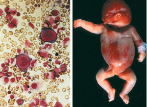
    Figure 5: B19-associated hydrops fetalis [55].
Panel A shows a bone marrow aspirate with no mature erythroid precursors and with characteristic giant pronormoblasts. In Panel B, hydrops fetalis is evident in
an infant who was infected in utero in midtrimester.
![]()
Authors (years)
Period of study
Country
Number of serum samples tested
Clinical Symptoms
Fetal Effects
[56]
1999-2004
Sendai, Japan
478

100 pregnant women had been exposed to B19 (21%)
49 51

Symptomatic Asymptomatic
Facial Rasch (51%) (25/49)
Most common Infection

Children living at home
Hydrops fetalis et fetal death
7% (7/100)
>20 weeks[57]
2005-2010
Bologna, Italy
72

68 pregnant women had been exposed to B19 (94.1%)
29 39

Symptomatic Asymptomatic
Rash, fever and polyarthralgia
(79.3%) 23/29-10,2% fetal deaths
-11,9 % Hydrops fetalis[58]
2004
Stuttgart, Germany
1018
-
- 6.3% fetal deaths (64/1018)
-3.9 % Hydrops fetalis (40/1018)[59]
1985-1988 and 1992-1995
London
420 pregnant women with B19 infection
-
Fetal loss averaged 9%
[60]
2010
Germany
236 pregnant women with B19 infection
-
-18.8% fetal loss(8/236)
- 23.6 % Hydrops fetalis (10/236)[61]
2014
Paris, France
-
-
20 cases of congenital parvovirus B19 Infection (1/20 (neonatal death)
Table 2: General characteristics of studies reporting on outcome of fetuses with parvovirus B19 (PB19) infection included in systematic review.
The vertical transmission of B19V occurs in about one third of women infected during pregnancy. Consequently, in the first trimester, the risk of fetal loss is 5-10%, while in the second trimester is about 11-12, 5%. It is considered that up to 20% nonimmune hydrops fetalis may be caused by B19V [54].
Epidemiological Studies
Infection with B19 is very common and cases of infection have been reported all over the world in all seasons. Parvovirus B19 is active worldwide with neither ethnical nor geographical boundaries, albeit with some regional differences [18].
Generally, seropositivity is lowest among young children, rises to around 50% at puberty [41]. And in-creases further at lower rates throughout adulthood. Seropositivity is correlated with age, a history of transfusion, and urban residency.
Worldwide, the prevalence of PVB19 in pregnant women is variable, generally between 60% and 80%, with lower prevalence in Asian regions [62].
Data on the seroprevalence of women susceptible to B19 infection in early pregnancy report a value of 26% to 43.5% in European countries and Japan [43,49,63].
In developing countries, the percentages are very similar, although the incidence and extent of infection have been studied in smaller population samples (Table 3).
![]()
Reference number
Year
Country
Total number of
serum samples testedNumber of serum
samples
infected by B19Percentage
B19
positive[64]
2016
Ardabil, Iran
350
242
69.10%
[65]
2014
Azerbaijan, Iran
86
65
75.60%
[66]
2014-2015
Mwanza, Tanzania
258
142
55%
[67]
2013
Ogbomoso, Nigeria
231
55
24%
[68]
2012
Sudan
147
73
49.70%
[69]
2011
Tunisie
404
307
76.20%
[70]
2007
Cordoba, Spain
42
27.7
66%
[71]
2007-2008
Tripoly, Libya
150
100
66%
[72]
2007-2008
Bialystok, Poland
55
24
43%
[73]
2008-2009
Khartoum state, Sudan
500
287
57.40%
[74]
2003-2010
Milan, Italy
37
29
78.30%
[75]
1999
Kuwait
1047
560
53.30%
[56]
1999-2004
Sendai, Japan
478
100
21%
[76]
1999-2008
Oslo, Norway
1349
832
61.70%
[77]
1998-2000
Nijmegen, The Netherlands
2567
1788
70%
Table 3: Prevalence of Parvovirus B19 infection among normal and at risk pregnant women.
Up to 2374 B19 infections occur annually in Japan among pregnant women. The risk of fetal death after infection is 9% during the first 20 weeks of pregnancy and up to 107 fetal deaths per year. Similarly, 2.9% risk of fetal hydrops between 9-20 weeks of pregnancy or 21 cases are estimated each year [63].
Laboratory Testing Options, Applications and Interpretation
Two circumstances lead to the diagnosis of parvovirus B19 infection in pregnant women: the occurrence of maternal clinical signs, mainly rash and arthritic manifestations, and the incidental finding of a fetoplacental hydrops [63]. The search for parvovirus B19 in the laboratory is based on a multipara metric approach, combining the immunological search for specific antibodies and the molecular detection of viral DNA [78].
Cell culture
The detection of viral particles in serum or bone marrow is not commonly used. In fact, PVB19 culture is only obtained by inoculating samples with fresh human marrow cells from a healthy donor. It is a cumbersome and expensive technique reserved for research [3].
Serology
A variety of methods can be used to detect parvovirus B19 antibodies (Table 4), and an international standard for B19 IgG assays has been developed and tested in collaborative studies [79,80]. Numerous diagnostic kits are available, most of which are based on enzyme immunoassay, immunofluorescence, or immunoblot techniques.
![]()
Manufacture
Methodc
Antigen and Source
FDA approved
Botrin International
(Dublin Ireland)Indirect IgG EIA and class-capture IgM EIA
Baculovirus-expressed VP2a
Yes
Diasorin (Saluggia, Italy)
Indirect IgG EIA and class-capture IgM CLIA-Liaison platform
Baculovirus-expressed VP2a
No
Denka Seiken (Tokyo, Japan)
Indirect IgG and IgM EIA
Baculovirus-expressed VP2a and VP2
No
Medac Diagnostika
(Wedel, Germany)Indirect IgG EIA and class-capture IgM EIA
Baculovirus-expressed VP2a and VP2
No
Euroimmun(LÜbeck,
Germany)Indirect IgG EIA and class-capture IgM EIA
yeast-expressed VP2
No
IBL(Hamburg, Germany)
Indirect IgG and IgM EIA
E.coli-expressed VP1
No
Focus Parvovirus Dxselect
(Cyprus, CA)Indirect IgG and IgM EIA
Recombinant VP1
No
Mikrogen (Martinsried, Germany)
Indirect IgG and IgM and Strip immunoassay
E.coli-expressed VP1
baculovirus-expressed VP2No
Botrin International
(Dublin Ireland)Indirect IgG and IgM immunofluorescebce (IFA)b
Baculovirus-expressed VP1
No
aVP2 comprises >95% of capsid antigen and appears to be conserved among genotypes.
bPretreatment with adsorbent reagent is needed to prevent interference from rheumatoid factors.
cEIA: Enzyme Immunoassay; CLIA: Chemiluminescence Immunoassay.
Table 4: Parvovirus B19 Serologic assays [80].
In general, IgM to B19 appears 7 to 10 days after infection, is followed with in a few days by IgG, and remains positive for 2 to 4 months. In contrast, immunocompromised hosts may not develop antibodies, or IgM can develop but remain positive for months or years as an indicator of persistent infection, without development of IgG. A positive IgM test will indicate primary infection. If IgM and IgG negative, the patient is at risk of primary infection and further tests are required if there is a strong clinical suspicion [81].
![]()
    Sample
    Technique


    Maternal serum
    IgG/IgM, ELISA, Western blot, immunofluorescence


    Maternal serum
    In situ hybridization


    Maternal serum
    AFP (elevated prior to detection of hydrops)


    Maternal serum
    AFP (elevated )


    Maternal serum
    PCR


    Maternal serum
    IgG antibodies against viral non-structural    protein NSI


    Amniotic fluid
    PCR


    Amniotic fluid
    PCR, Southern blot, chemiluminescence


    Amniotic fluid
    Enzymatic amplification of parvovirus B19 (segment)


    Fetal blood
    PCR, dot-blot hybridization, in situ hybridization


    Fetal blood
    Enzymatic amplification of parvovirus B19 (segment)


    Fetal serum
    PCR


    Fetal heart tissue
    Microscopy: VP1 and VP2 viral particles


    Fetal tissue
    Microscopy: histology was found to be as    sensitive as PCR


    Ig: Immunoglobulin; ELISA: Enzyme-Linked    Immunosorbent Assay; AFP: Alpha-Fetoprotein; PCR: Polymerase Chain Reaction    (DNA test).
Table 5: Diagnostic techniques and samples used for diagnosing congenital
parvovirus B19 infections [87].
When only IgG is positive, the doubt may persist if the antibody test was performed at a distance from the clinical point of call or the contact person: if the levels are low and stable, it is certainly an old infection; if the levels are high or have ascending kinetics, it may be a re-infection or a primary infection with IgM that has already disappeared from the serum [82].
Viral DNA detection (PCR)
PCR techniques to quantify viral DNA are commonly used. The most widely used technique is real-time PCR, which gives very rapid results with very high sensitivity [3].
The search for DNA in maternal blood can sometimes help in diagnosis because PCR is positive in the first month following primary infection; DNA then decreases progressively. This PCR can also be performed on abortion products or fetal tissue taken during an autopsy. A preliminary study has shown that amniotic fluid samples are much richer in virus than maternal blood collected at the same time [83]. In the case of low IgM and viral DNA levels with a strong suspicion of contagion, it is possible to test for antibodies to VP2 and VP1, thus allowing a very early diagnosis of seroconversion and thus the implementation of fetal surveillance [84].
PCR also appears to be of particular interest for the analysis of MFIU in the first and third trimesters of unknown etiology [85,86].
Anatomopathological analysis
This analysis allows the detection of intranuclear viral inclusions and margined chromatin, particularly in erythroid progenitors (lungs, liver, thymus, kidneys, etc.). PVB19 is suspected to cause malformations in the fetus, but very few cases are reported and they only concern spontaneous abortions. Ocular anomalies, myocardial necrosis, and intestinal vascular accidents have been described [88].
Ultrasound examination of the fetus
Ultrasound monitoring, by evaluating the degree of hydrops and performing Doppler analysis of the systolic peak of the middle cerebral artery, will allow the severity of fetoplacental hydrops to be assessed. It will make it possible to distinguish between two types of anasarca: early anasarca and pericardial anasarca [89].
Antenatal biological diagnosis
Prenatal diagnosis of fetal B19 infection is made by testing for the virus or viral genome in amniotic fluid (PCR) or fetal blood.
B19 infection during pregnancy can affect the fetus and result in hydrops or fetal death. Anasarca is the predominant ultrasound feature in fetuses with parvovirus B19 infection. Obtaining a diagnosis of maternal infection will allow fetal evaluation and treatment by intrauterine blood transfusion. Unfortunately, mothers are often unaware that they have an infection until fetal symptoms are noted. Confirmation of PVB infection19 requires laboratory analysis, which is complicated by the nature of the viral infection and the immune response [90].
Management of Infection to B19
After contact, surveillance at any term is maintained for 12 weeks because there is considered to be a 10% risk of complications before 28SA that drops to less than 1% but still exists after 28SA. A latency of 6 weeks on average between maternal infection and the appearance of fetoplacental anasarca is also noted in the literature [91].In case of contagion, maternal serology should be checked to determine the initial status and recheck 2 to 3 weeks later (Figure 6).
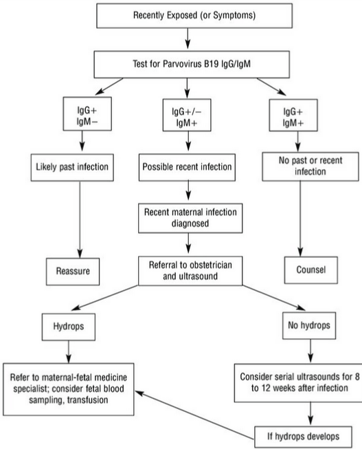
Figure 6: Management of a pregnant woman exposed to parvovirus B19 infection [92].
Prognosis, Prevention and Therapeutic Approaches
Prognosis
Several studies have shown that the short- and long-term prognosis of children born alive to mothers infected with PVB19 is excellent, with 98% of children surviving without sequelae, even if the mother is infected, in only 17 to 33% of cases [59,93].
The prognosis is clouded by spontaneous abortions and hydrops. The prognosis is clouded by spontaneous abortion and anasarca. Indeed, the prognosis of fetuses in anasarca having been transfused in utero is considered favorable in the light of several studies [94,95].
Prevention
Mothers of young children, as well as women working in schools and daycare centers, are most at risk of exposure to B19 infection. Excluding infected people from the workplace, daycare or school is not likely to prevent the spread because infected people are contagious before symptoms appear [58].
To reduce the risk of infection, pregnant women should:
- Wash their hands thoroughly after touching tissues used by infected children and dispose of tissues immediately.
- Avoid sharing glasses or utensils with anyone who has or has been exposed to the disease.
No systematic prevention or screening methods are effective.
Therapeutic approaches
There is no specific antiviral treatment for PVB19. The primary infection of the immunocompetent subject generally does not require any specific treatment. In patients with chronic hemolysis, transfusion of packed blood cells and injection of polyvalent immunoglobulins may be warranted.
Intrauterine blood transfusion has been proposed as a treatment for the fetus with severe B19-induced anemia and hydrops. However, the natural history of infection in the untreated fetus is not known, and the benefit of intrauterine blood transfusion is as yet unproven. Several investigators have reported spontaneous resolution of nonimmune hydrops. A survey of perinatal obstetricians in the United States and Canada reported that 34% of the cases of nonimmune hydrops resulting from parvovirus infection resolved spontaneously, the majority within 8weeks [96]. In the absence of such treatment, fetal death occurs in 90% of cases [97].
B19 vaccine
As far as is currently known, no vaccine is currently available. A Phase I study working on a recombinant vaccine comprising the capsid proteins VP1 and VP2 seems to confirm the good immunogenicity of these proteins inducing the production of a high level of antibodies persisting at least one year [98].
References
- Cohen BJ. Human parvovirus B19 and fifth disease. In: Mortimer PP, Public Health Virology 12 Reports 1st edn. London: Public Health Laboratory Service. 1986; 130-143.
- White DG, Woolf AD, Mortimer PP, Cohen BJ, Blake DR, Bacon PA. Human parvovirus arthropathy. Lancet. 1985; 1: 419-421.
- Heegaard ED, Brown KE. Human parvovirus B19.Clin Microbiol Rev. 2002; 15: 485-505.
- Lunardi C, Tinazzi E, Bason C, et al. Human parvovirus B19 infection and autoimmunity. Autoimmun Rev. 2008; 8: 116-120.
- Ozawa K, Young N. Characterisation of capsid and noncapsid proteins of B19 parvovirus propagated in human erythroid bone marrow cell cultures. Journal of virology. 1987; 61: 2627-2630.
- Parvovirus B19 (erythema infectiosum, fifth disease). In: Red Book 2006: Report of the Committee on Infectious Diseases. 27th ed. Washington, D.C.: American Academy of Pediatrics. 2006: 484-487.
- Enders M, Weidner A, Zoellner I, et al. Fetal morbidity and mortality after acute humane parvovirus B19 infection in pregnancy: prospective evaluation of 1018 cases. Prenat Diagn. 2004; 24: 513-518.
- Torok TJ. Parvovirus B19 and human disease. Adv Intern Med. 1992; 37: 431-455.
- Brochot C, Debever P, Subtil D, Puech F. Which supervision and treatment in case of exposure to Parvovirus B19 during pregnancy? Obstetrics and Fertility Gynecology. 2008; 36: 204-211.
- Yi-quan X, et al. The risk of maternal parvovirus B19 infection during pregnancy on fetal loss and fetal hydrops:A systematic review and metaanalysis. Journal of Clinical Virology. 2019; 114: 12-20.
- Labau E, Bonnet E, Alric L, Massip P. Parvovirus B19 infection. New pathophysiological approaches. Press Med 1996; 25: 162-166.
- Anderson JL, Young NS, Erdman DD. Human parvovirus B19. Laboratory diagnosis. In: Monographs in virology: human Parvovirus B19. Eds, Karger. Basel. 1997; 93-104.
- Zerbini M, Gallinella G, Cricca M, Bonvicini F, Musiani M. Diagnostic procedures in B19 infection. Pathol Biol (Paris). 2002; 50: 332-338.
- Cossart Y, Field AM, Cant B, Widdows D. Parvovirus-like particles in human sera. Lancet. 1975; 1: 72-73.
- Morinet F, Tchernia G. parvovirus B19 and hematopoiesis. Medecine- Sciences. 1991; 7: 127-137.
- Broliden K, Tolvenstam T, Norbeck O. Clinical aspects of parvovirus B19 infection. Journal of Internal Medicine. 2006; 260: 285-304.
- International Committee on Taxonomy of Viruses. Virus taxonomy: classification and nomenclature of viruses. Seventh report of the International Committee on Taxonomy of Viruses. Springer-Verlag, Vienna, Austria. 2000.
- Rogo LD, Mokhtari-Azad T, Kabir MH, Rezaei F. Human Parvovirus B19: A review. ActaVirologica. 2014; 58: 199-213.
- Kajigaya S, Fujii H, Field A, et al. Self-assembled B19 parvovirus capsids, produced in a baculovirus system, are antigenically and immunogenically similar to native virions. Proc Natl Acad Sci USA. 1991; 88: 4646-4650.
- Schwarz TF, Serke S, Von Brunn A, et al. Heat stability of Parvovirus B19: kinetics of inactivation. Zentralbl Bakteriol. 1992; 277: 219-223.
- Moffatt S, Yaegashi N, Tada K, Tanaka N, Sugamura K. Human parvovirus B19 nonstructural (NS1) protein induces apoptosis in erythroid lineage cells. J Virol. 1998; 72: 3018-3028.
- Chen Z, W Guan, F Cheng, AY Chen, J Qiu. Molecularcharacterization of human parvovirus B19 genotypes 2 and 3. Virology. 2009; 394: 276-285.
- W Guan, F Cheng, Y Yoto, S Kleiboeker, S Wong, N Zhi, et al. Block to the production of full-length B19 virus tran-scripts by internal polyadenylation is overcome by replication of the viral genome. J. Virol. 2008; 82: 9951-9963.
- S Pillet, N Le Guyader, T Hofer, F NguyenKhac, M Koken, JT Aubin, et al. Hypoxia enhanceshuman B19 erythrovirus gene expression in primary erythroid cells. Virol-ogy. 2004; 327: 1-7.
- Tewary SK, Zhao H, Deng X, Qiu J, Tang L. The human parvovirus B19 non-structural protein 1 N-terminal domain specifically binds to the origin of replication in the viral DNA. Virology. 2014; 449: 297-303.
- Young NS, Brown KE. Parvovirus B19. N Engl J Med. 2004; 350: 586-597.
- Erdman DD, Durigon EL, Holloway BP. Detection of human parvovirus B19 DNA PCR products by RNA probe hybridization enzyme immunoassay. J Clin Microbiol. 1994; 32: 2295-2298.
- Gallinella G, Venturoli S, Manaresi E, Musiani M, Zerbini M. B19 virus genome diversity: epidemiological and clinical correlations. J Clin Virol. 2003; 28: 1-13.
- Eis-Hubinger AM, Reber U, Edelmann A, Kalus U, Hofmann J. Parvovirus B19 genotype 2 in blood donations. Transfusion. 2014; 54: 1682-1684.
- Von Kietzell K, Pozzuto T, Heilbronn R, Grössl T, Fechner H, et al. Antibodymediated enhancement of parvovirus B19 uptake into endothelial cells mediated by a receptor for complement factor C1q.J. Virol. 2014; 88: 8102- 8115.
- Brown KE, Anderson SM, Young NS. Erythrocyte P antigen: cellular receptor for B19 parvovirus. Science. 1993; 262: 114-117.
- Bültmann BD, Klingel K, Sotlar K, Bock CT, Kandolf R. Parvovirus B19: a pathogen responsible for more than hematologic disorders. Virchows Arch. 2003; 442: 8-17.
- Koehl B, Oualha M, Lesage F, Rambaud C, Canioni D, et al. Fatal parvovirus B19 myocarditis in children and possible dysimmune mechanism. Pediatr Infect Dis J. 2012; 31: 418-421.
- Peterlana D, Puccetti A, Corrocher R, Lunardi C. Serologic and molecular detection of human parovirus B19 infection. Clin Chim Acta. 2006; 372: 14- 23.
- LJ Anderson, C Tsou, RA Parker, TL Chorba, H Wulff, P Tattersall, et al. Detection of antibodies and antigens of human parvovirus B19 by enzymelinked immunosorbent assay. J. Clin. Microbiol. 1986; 24: 522-526.
- DD Erdman, MJ Usher, C Tsou, EO Caul, GW Gary, S Kajigaya, et al. Human parvovirus B19 specific IgG, IgA, and IgM antibodies and DNA in serum specimens from persons with erythema infectiosum. J. Med. Virol. 1991; 35: 110-115.
- MJ Anderson, PG Higgins, LR Davis, JS Willman, SE Jones, IM Kidd, et al. Experimental parvoviralinfection in humans. J. Infect. Dis. 1985; 152: 257- 265.
- Magro CM, Dawood MR, Crowson AN. The cutaneous manifestations of human parvovirus B19 infection. Hum Pathol. 2000; 31: 488-497.
- Corman LC, Dolson DJ. Polyarteritis nodosa and parvovirus B19 infection. Lancet. 1992; 339: 491.
- Broliden K, Tolfvenstam T, Norbeck O. Clinical aspects of parvovirus B19 infection. Journal of Internal Medicine. 2006; 260: 285-304.
- Mossong J, Hens N, Friederichs V, Davidkin I, Broman M, Litwinska B, et al. Parvovirus B19 infection in five European countries: seroepidemiology, force of infection and maternal risk of infection. Epidemiol Infect. 2008; 136: 1059-1068.
- Ergaz Z, Ornoy A. Parvovirus B19 in pregnancy. Reprod Toxicol. 2006; 21: 421-435.
- Dijkmans AC, de Jong EP, Dijkmans BA, Lopriore E, Vossen A, Walther FJ, et al. Parvovirus B19 in pregnancy: prenatal diagnosis and management of fetal complications. Curr Opin Obstet Gynecol. 2012; 24: 95-101.
- Lamont RF, Sobel JD, Vaisbuch E, Kusanovic JP, Mazaki-Tovi S, Kim SK, et al. Parvovirus B19 infection in human pregnancy. BJOG. 2011; 118: 175-186.
- Valeur-Jensen AK, Pedersen CB, Westergaard T, Jensen IP, Lebech M, Andersen PK, et al. Risk factors for parvovirus B19 infection in pregnancy. JAMA. 1999; 281: 1099-1105.
- de Jong EP, Walther FJ, Kroes AC, Oepkes D. Parvovirus B19 infection in pregnancy: new insights and management. Prenat Diagn. 2011; 31: 419-425.
- Chorba T, Coccia P, Holman RC, Tattersall P, Anderson LJ, Sudman J, et al. The role of parvovirus B19 in aplastic crisis and erythema infectiosum (fifth disease). J Infect Dis. 1986; 154: 383-393.
- Rodis JF, Hovick TJ Jr, Quinn DL, Rosengren SS, Tattersall P. Human parvovirus infection in pregnancy. Obstet Gynecol 1988; 72: 733-738.
- Jensen IP, Thorsen P, Jeune B, Moller BR, Vestergaard BF. An epidemic of parvovirus B19 in a population of 3,596 pregnant women: a study of sociodemographic and medical risk factors. BJOG. 2000; 107: 637-643.
- Koch WC, Harger JH, Barnstein B, Adler SP. Serologic and virolologic evidence for frequent intrauterine transmission of human parvovirus B19 with a primary maternal infection during pregnancy. Pediatric Infect Dis J. 1998; 17: 489-494.
- Bonvicini F, Bua G, Gallinella G. Parvovirus B19 infection in pregnancyawareness and opportunities. Curr Opin Virol. 2017; 27: 8-14.
- Young NS, Brown KE. Parvovirus B19. N Engel J Med. 2004; 350: 586-597.
- Brown KE. Parvovirus B19. In: Mandell, Douglas, and Bennett’s Principals and Pratice of infections Diseases. Eds, Mandell GL, Bennett JE, Dolin R. 6th ed. Churchil Livingstone Elsevier. Philadelphia, PA. 2005; 2: 1981-1902.
- Brown T, Anand A, Ritchie LD, et al. intrauterine parvovirus infection associated with hydrops fetalis. Lancet. 1984; 2: 1033-1034.
- Neal S Young, Kevin E Brown. Parvovirus B19. N Engl J Med. 2004; 350: 586-597.
- Chisaka H, Ito K, Niikura H, Sugawara J, Takano T, Murakami T, et al. Clinical Manifestations and outcomes of parvovirus B19 infection during pregnancy in Japan. Tohoku J.Exp.Med. 2006; 209: 277-283.
- Bonvicini F, Puccetti C, Salfi NCM, Guerra B, Gallinella G, Rizzo N, et al. Gestational and Fetal Outcomes in B19 Maternal Infection: a Problem of Diagnosis. Journal of clinical microbiology. 2011; 49: 3514-3518.
- Enders M, Weidner A, Zoellner I, Searle K, Enders G. Fetal morbidity and mortality after acute human parvovirus B19 infection in pregnancy: prospective evaluation of 1018 cases. Prenat Diagn. 2004; 24: 513-518.
- E Miller, CK Fairley, BJ Cohen, C Seng. Immediate and long term outcome of human parvovirus B19 infection in pregnancy. Br J Obstet Gynaecol. 1998; 105: 174-178.
- Enders M, Karin Klingel, Andrea Weidner, Carola Baisch, Reinhard Kandolf, Gunnar Schalasta, et al. Risk of fetal hydrops and non-hydropic late intrauterine fetal death after gestational parvovirus B19 infection. Journal of Clinical Virology. 2010; 49: 163-168.
- Macé G, Marine Sauvan, Vanina Castaigne, Marie-Laure Moutard, Anne Cortey, et al. Clinical presentation and outcome of 20 fetuses with parvovirus B19 infection complicated by severe anemia and/or fetal hydrops. Prenatal Diagnosis. 2014; 34: 1023-1030.
- Morinet F, Pallier C, Pillet S, Huraux JM, Nicolas JC, Augut H, Peiguelafeuille H Parvoviridae. Estem (eds) traité de virologie médicale, Paris. 2003; 283-289.
- Nabae K, Satoh H, Nishiura H, Tanaka-Taya K, Okabe N, Oishi K, et al. Estimating the risk of parvovirus B19 infection in blood donors and pregnant women in Japan. Plos one. 2014; 9: e92519.
- Habibzadeh S, Peeri-Doghaheh H, Mohammad-Shahi J, Mobini Elham, Shahbazzadegan S. The prevalence of parvovirus B19 infection among pregnant women of Ardabil in 2013. Iranian journal of Microbiology. 2016; 8: 214-218.
- ZR Khameneh, H Hanifian, R Barzegari, N Sepehrvand. Human parvovirus B19 in Iranian pregnant women: A serologic survey. indian journal of pathology and microbiology. 2014; 57: 442-444.
- Mariam M. Mirambo, Fatma Maliki, Mtebe Majigo, Martha F Mushi, Nyambura Moremi, Jeremiah Seni, Dismas Matovelo and Stephen E. Mshana. The magnitude and correlates of Parvovirus B19 infection among pregnant women attending antenatal clinics in Mwanza, Tanzania. BMC Pregnancy and Childbirth 2017; 17: 176.
- Iyanda Abiodun, Oluyinka Oladele Opaleye, Olusola Ojurongbe, Ademola Hezekiah Fagbami. Seroprevalence of parvovirus B19 IgG and IgM antibodies among pregnant women in Oyo State, Nigeria. J Infect Dev Ctries. 2013; 7: 946-950.
- Gasim I Gasim, Reem Eltayeb, Elhassan M Elhassan, AbdElrahium D Haggaz, Duria A Rayis, Ishag Adam. Human parvovirus B19 and low hemoglobin levels in pregnant Sudanese women. Int J Gynaecol Obstet. 2016; 132: 318-320.
- N Hannachi, M Marzouk, I Harrabi, A Ferjani, Z Ksouri, H Ghannem, et al. Seroprevalence of rubella virus, varicella zoster virus, cytomegalovirus and parvovirus B19 among pregnant women in the Sousse region, Tunisia. Bulletin de la Société de pathologie exotique. 2011; 104: 62-67.
- MS Pedranti, MP Adamo, R Macedo, MT Zapata. Prevalence of anti-rubella and anti-parvovirus B19 antibodies in pregnant women in the city of Córdoba, and in women of fertile age in the city of Villa Mercedes, province of San Luis. Rev Argent Microbiol. 2007; 39: 47-50.
- Elfatah Elnifro, AK Nisha, Musbah Almabsoot, Ali Daeki, Nuri Mujber, Jose Muscat. Seroprevalence of parvovirus B19 among pregnant women in Tripoli, Libya. J Infect Dev Ctries. 2009; 3: 218-220.
- Agata Zajkowska, Adam Garkowski, Piotr Czupryna, Anna Moniuszko, Monika Emilia Król, Jacek Szamatowicz, et al. Seroprevalence of parvovirus B19 antibodies among young pregnant women or planning pregnancy, tested for toxoplasmosis. Przegl Epidemiol. 2015; 69: 479-82, 597-600.
- O Adam, T Makkawi, U Reber, H Kirberg, AM Eis-Hübinger. The seroprevalence of parvovirus B19 infection in pregnant women in Sudan. Epidemiol Infect. 2015; 143: 242-248.
- Maurizio Zavattoni, Stefano Paolucci, Antonella Sarasini, Beatrice Tassis, Mariangela Rustico, Aida Quarenghi, et al. Diagnostic and prognostic value of molecular and serological investigation of human parvovirus B19 infection during pregnancy. New Microbiol. 2016; 39: 181-185.
- Ma’asoumah Makhseed, Alexander Pacsa, Mohammad Abrar Ahmed, Sahar Sultan Essa. Pattern of Parvovirus B 19 Infection during Different Trimesters of Pregnancy in Kuwait. Infectious Diseases in Obstetrics and Gynecology. 1999; 7: 287-292.
- Barlinna, Halvor Rollagb, Lill Trogstada, Kirsti Vainioa, Coraline Basseta, Per Magnusc, et al. Dudmana High incidence of maternal parvovirus B19 infection in a large unselected population-based pregnancy cohort in Norway. Journal of clinical virology. 2017; 94: 57-62.
- Peter H van Gessel, Michael A Gaytant, Ann CTM Vossen, Joep MD Galama, Nicolette TC Ursem, Eric AP Steegers, et al. Incidence of parvovirus B19 infection among an unselected population of pregnant women in the Netherlands: A prospective study. Eur J Obstet Gynecol Reprod Biol. 2006; 128: 46-49.
- Gallinella G. Molecular testing for Parvovirus. In Diagnostic Molecular Pathology. Edited by Coleman WB, Tsongalis GJ. Academic Press. 2017; 103-113.
- Ferguson M, Heath A. Report of a collaborative study to calibrate the Second International Standard for parvovirus B19 antibody. Biologicals. 2004; 32: 207-212.
- Landry ML. Parvovirus B19. Microbilogy Spectrum. 2021; 4: 5.
- Crane J, et al. Infection au parvovirus B19 encours de grossesse. JSOGS. 2002; 119.
- de Jong EP, et al. Parvovirus B19 infection in pregnancy. Journal of clinical virology. 2006; 36: 1-7.
- Knoll A, Louwen F, Kochanauski B, Plentz A, Stussel J, Beckenlehner K, et al. Parvovirus B19 infection in pregnancy: quantitative viral ADN analysis using a kinetic flurescence detection system (Taq Man PCR), J. Med. Virol. 2002; 67: 259-266.
- Enders M, Schalasta G, Baisch C, et al. Human parvovirus B19 infection during pregnancy value of modern molecular and serological diagnostics. J. Clin. Virol. 2006; 35: 400-406.
- Tolfvenstam T, Papadogiannakis N, Norbeck O, Petersson K, Broliden K. Frequency of human parvovirus B19 infection in intrauterine fetal death. Lancet. 2001; 357: 1494-1497.
- Skjoldebrand-Sparre L, Tolfvenstam T, Papadogiannakis N, Wahren B, Broliden K, Nyman M. Parvovirus B19 infection: association with third trimester intrauterine fetal death. Br J Obstet Gynaecol. 2000; 107: 476-80.
- Von Kaisenberg CS, Jonat W. Fetal parvovirus B19 infection. Ultrasound Obstet Gynecol. 2001; 18: 280-288.
- Brown KE. What threat is human parvovirus B19 to the fetus? Br J Obstet Gynaecol. 1989; 96: 764-767.
- Benoist G, Dina J, Herlicoviez M. Le diagnostic prénatal en pratique. 2011; 339-344.
- Crane J, Mundle W, Boucoiran I. Infection au parvovirus B19 pendant la grossesse. 2014; 36: 1117-1118.
- Subtil D, Garabedian C, Chauvet A. Infection à Parvovirus B19 et grossesse. La presse médicale. 2015; 44: 13.
- Faden Y. Toxoplasmosis, Cytomegalovirus, Herpes Simplex Virus, Rubella, Parvovirus, and Listeria Infections. In: Medical Care of the Pregnant Patien. 2nd edition. Lee R, Keely E, Rosella-Montella K. American College of Physicians. 2007; 55: 687-708.
- Gratacos E, et al. The incidence of human parvovirus B19 infection during pregnancy and its impact on perinatal outcome. J Infect Dis. 1995; 171: 1360- 1363.
- Dembinski J, Haverkamp F, Maara H, Hansmann M, Eis-Hubinger AM, Bartmann P. Neurodevelopmental outcome after intrauterine red cell transfusion for parvovirus B19 - induced fetal hydrops. Bjog. 2002; 109: 1232- 1234.
- Vanspranghels R, Houfflin-Debarge V, Vaast P, Coulon C, Clouqueur E, Hanssens S, et al. Transfusion. 2019; 59: 185-190.
- Rodis JF, Borgida AF, Wilson M, Egan JFX, Leo MV, Odibo AO, et al. Management of parvovirusinfection in pregnancy and outcomes of hydrops: A surveyof members of the Society of Perinatal Obstetricians. American Journal of Obstetrics and Gynecology. 1998; 179: 985-988.
- Bousquet F, Segondy M, Faure JM, Deschamps F, Boulot P. B19 parvovirus induced fetal hydrops : good outcome after intrauterine blood transfusion at 18 weeks of gestation. Fetal Diagn Ther. 2000; 15: 132-133.
- Ballou WR, Reed JL, Noble W, Young NS, Koenig S. Safety and immunogenicity of a recombinant parvovirus B19 vaccine formulated with MF59C. 1. J Infect Dis. 2003; 187: 675-678.