
Abstract
A 55-year-old man with a history of melanoma in situ, renal insufficiency, COPD, and smoking history presented with an 8-month history of worsening pain, erythema, edema, and progressive deformity of the right fifth finger. An X- ray revealed a mass with obliteration of bony structures. The near absence of the proximal and distal phalanx on imaging was highly suspicious for a metastatic process. A biopsy was performed which demonstrated changes consistent with tophaceous gout. Chronic tophaceous gout generally arises due to prolonged accumulation of u rate crystals which result in destruction of joints and surrounding tissue. Occasionally this accumulation can be more abrupt and lead to bony destruction which can initially be mistaken for metastatic disease. Primary malignancies that commonly metastasize to the bone include lung, breast, and prostate, melanoma, thyroid and renal. The overlapping clinical and radiographic findings can be particularly concerning in patients with a pre-existing cancer history. We present a patient with an unusual initial presentation of acute gout that arose with complete obliteration of the distal phalanx of the 5th digit as initial presentation of disease.
Case Presentation
A 66-year-old man with a history of melanoma in situ, alcohol abuse, COPD, renal insufficiency, and prior smoking with previous normal CT lung screening presented with an 8-month history of worsening pain, erythema, edema, and deformity of the 5 digits on the right hand. He reports that the nail under the digit has begun to cause him discomfort and notes the appearance of drainage when he clips the nail or squeezes the finger.
Physical exam revealed a trumpet deformity of the nail on the right 5th digit. The finger was warm to touch on both sides of the loosely adherent nail. While mobility was not compromised, he did endorse pain in the right DIP of the small finger. X- Ray of the right hand revealed a mass on the AP lateral projections of the right small finger with obliteration of bony structures. The near absence of the proximal and distal phalanx on imaging was highly suspicious for bone metastasis. A complete blood count was ordered at time of visit noted only mild anemia (hemoglobin of 12.9). A complete metabolic panel was within normal limits expecting for glucose of 116 and blood urea nitrogen of 22. A biopsy was performed. The biopsy specimen displayed bone fragments and a palisading histiocytic reaction surrounding polarizable crystal-like material. These findings are consistent with gout and there was no evidence of malignancy seen. The patient opted to have the distal part of digit removed and improvement of his pain was noted at follow-up.
Discussion
Gout is a common inflammatory arthropathy in adults that results from an inflammatory reaction to urate deposition in joints and tissues. The condition mostly effects middle-aged to elderly men and postmenopausal women. The prevalence of gout falls between 1% to 6.8% of the general population worldwide [1]. Risk factors for gout include consumption of purine-rich foods, alcoholic beverages, metabolic syndrome, hypertension, family history, and chronic renal disease. In addition, Exposure to chemotherapeutic regimens can lead to elevated uric acid levels secondary to lysis of tumor cells. Acute disease typically presents on the lower extremities, but eventually spreads to the joints and axial areas. When untreated this can result in renal failure, and accumulation of urate crystals can cause painfully, destructive bone erosions, as seen in this case.
While most cases of gout are diagnosed clinically the diagnosis can be confirmed with observation of monosodium urate crystals on biopsy [2,3]. Of note, the crystals do not fix well in formalin and for best visualization should be fixed in alcohol [3]. Plain radiographic images are usually normal at initial presentation [4,5]. However, late manifestations result in tophi which are seen as intraosseous masses associated with well-defined erosive arthropathy, replacement or widening of joint spaces and subchondral collapse [4,5]. This case describes an uncommon initial presentation of erosive gout as bone destruction initially concerning for metastasis.
Tophi are classic indication of chronic gout. Tophi usually form after years of uncontrolled gout but the exact mechanism behind formation has yet to fully elucidate. Tophi occur in about 12% to 35% of gout patients and are easily preventable with adequate control of the disease with uric acid lowering agents [6]. Bone erosion is a frequent manifestation of chronic tophaceous gout that can lead to joint damage and deformity and, eventually, musculoskeletal disability. There is a strong association between tophus gout and bone erosion as studies have shown monosodium urate crystals in subchondral bone indicating direct contact of bone cells with the crystals [7]. Furthermore, another study has shown monosodium urate crystals reduce the function of osteoblast viability which is important for bone formation [8].
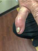
    Figure 1: Right 5th digit noted trumpet type deformity of the nail with loose
adherence and erythema and edema of the distal interphalangeal bone.
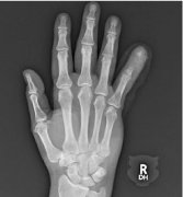
    Figure 2: AP lateral x-ray showing projections of the right small finger with
demonstrations of obliteration of bony structures.
Gouty arthritis may mimic bone metastasis. Primary care physicians and medical oncologists should be aware of the similarities that can be seen both clinically and on imaging. Primary malignancies that commonly metastasize to the bone include lung, breast, and prostate, melanoma, thyroid and renal. However, the digits are a rare site for metastasis. When this occurs, they are typically bronchogenic in origin and associated with very poor outcomes [9]. Lung cancer is the most common cause of acrometastasis in the hand and can be the initial presentation of occult malignancy [10]. Radiographic findings for metastases often depict lytic bone lesions with associated thinning of the trabeculae and ill-defined margins that indicate regions of destroyed trabeculae between the central destruction and radiological normal bone (similar to the findings in our patient) [11]. A biopsy is inexpensive and informative tests that can differentiate between the two as both the psychological and prognostic significance differ greatly.
Given our patient’s history and the onset of presentation one can also include the disease Multicentric Reticulo Hyistocytosis (MRH) in the differential. MRH is a rare inflammatory disease with cutaneous lesions and an associated erosive aggressive arthritis. Classically, variably sized brown-red or flesh-colored papules and nodules are found in linear arrangement on periungual skin. Some refer to this distribution as the “coral bead sign” [12]. Radiographic findings commonly show severe destruction of the distal interphalangeal joints. This pattern of bone erosions with sclerotic margins and overhanging edges may mimic findings commonly seen in gout [12]. MRH has been reported as an associated paraneoplastic manifestation in up to 25% of malignancies [12].
Treatment of gout focuses on prevention of reoccurring episodes and symptom relief of pain and inflammation. Colchicine, nonsteroidal anti-inflammatory agents and steroids are used to control acute attacks. Long term control is maintained using urate-reducing agents like probenecid, allopurinol, or rasburicase and adequate hydration [1]. The choice of agents used is dependent on the patient and the comorbidities they possess. Surgical removal of affected area is warranted for symptomatic relief after failed medical management.
Conclusion
In summary, gout is important to consider in the differential diagnosis of bone metastasis when dealing with painful lytic lesions on the hands as treatment and prognosis differ greatly between the two. It is important for clinicians to consider the possibility of an atypical presentation of gout in patients with painful lytic lesions given the differences in prognosis and management. This is especially relevant in patients with renal insufficiency, those with risk factors for malignancy development, and those on chemotherapeutics as they may develop more accelerated disease secondary to decreased renal clearance of uric acid.
References
- Dehlin M, Jacobsson L, Roddy E. Global epidemiology of gout: prevalence, incidence, treatment patterns and risk factors. Nat Rev Rheumatol. 2020; 16: 380-390.
- Parathithasan N, Lee WK, Pianta M, Oon S, Perera W. Gouty arthropathy: Review of clinico-pathologic and imaging features. J Med Imaging Radiat Oncol. 2016; 60: 9-20.
- Ragab G, Elshahaly M, Bardin T. Gout: An old disease in new perspective-A review. J Adv Res. 2017; 8: 495-511.
- McQueen FM, Reeves Q, Dalbeth N. New insights into an old disease: advanced imaging in the diagnosis and management of gout. Postgrad Med J. 2013; 89: 87-93.
- Perez-Ruiz F, Dalbeth N, Urresola A, de Miguel E, Schlesinger N. Gout. Imaging of gout: findings and utility. Arthritis Res Therapy. 2009; 11: 1-8.
- Kasper IR, Juriga MD, Giurini JM, Shmerling RH. Treatment of tophaceous gout: when medication is not enough. Semin Arthritis Rheum. 2016; 45: 669-674.
- Dalbeth N, Clark B, Gregory K, Gamble G, Sheehan T, Doyle A, et al. Mechanisms of bone erosion in gout: a quantitative analysis using plain radiography and computed tomography. Ann Rheum Dis. 2009; 68: 1290-1295.
- Chhana A, Callon KE, Pool B, Naot D, Watson M, Gamble GD, et al. Monosodium urate monohydrate crystals inhibit osteoblast viability and function: implications for development of bone erosion in gout. Ann Rheum Dis. 2011; 70: 1684-1691.
- Cho YJ, Cho YM, Kim SH, Shin K-H, Jung S-T, Kim HS. Clinical analysis of patients with skeletal metastasis of lung cancer. BMC cancer. 2019; 19: 303.
- Spiteri V, Bibra A, Ashwood N, Cobb J. Managing acrometastases treatment strategy with a case illustration. Ann R Coll Surg Engl. 2008; 90: W8-11.
- Milovanovic IS, Stjepanovic M, Mitrovic D. Distribution patterns of the metastases of the lung carcinoma in relation to histological type of the primary tumor: An autopsy study. Ann Thorac Med. 2017; 12: 191-198.
- Tajirian AL, Malik MK, Robinson-Bostom L, Lally EV. Multicentric reticulohistiocytosis. Clin Dermatol. 2006; 24: 486-492.