
Abstract
Multiple Sclerosis (MS) can present diagnostic challenges to clinicians due to its varied clinical presentations and lack of specific biomarkers. The McDonald criteria can aid in diagnosis but may require serial MRI scans to detect focal white matter lesions. Primary care clinicians are likely to encounter patients with new onset neurologic symptoms suggestive of MS or chronic, nonspecific, neurologic symptoms without a clear diagnosis. Long-term follow-up, re-evaluations and serial testing is a strong component of the model of care provided by primary care clinicians which can enable earlier diagnosis and access to comprehensive treatment. Clinicians must be vigilant to avoid premature closure by anchoring on to a diagnosis based on insufficient criteria. This report describes a patient with an atypical case of relapsing-remitting MS which went undiagnosed for a decade despite periodic evaluations by different clinicians, CNS imaging and specialist consultation. Following diagnosis, the patient was started on disease modifying therapy and engaged more comprehensively in the overall care of the physical and behavioral aspects of MS with measurable improvement.
Keywords: Multiple Sclerosis; Demyelination; Magnetic resonance imaging
Case Presentation
A 40-year-old African-American woman with a past medical history of asthma, sickle cell trait, B12 deficiency, morbid obesity and knee osteoarthritis presented to the family medicine clinic with a two-week history of diffuse paresthesia, weakness and associated body pain with centripetal radiation and sparing of the face, neck and shoulders. The patient reported severe physical dysfunction and inability to work due to difficulty with gait, lower extremity coordination and pain radiating distally from the thighs while walking. She also reported a metallic taste in the mouth. Further, the patient endorsed recent onset of mild urinary incontinence and depressed mood. There were no visual changes, heat sensitivity, skin rash or diurnal variation in symptoms. A cursory evaluation at an urgent care facility at the time of symptom onset revealed mild microcytic anemia, dehydration, normal chest x-ray and unremarkable 12-lead EKG.
The patient reported experiencing similar, brief, self-resolving and intermittent episodes of diffuse pain and paresthesia of lesser intensity over the past ten years. These were previously attributed to chronic pain syndrome and fibromyalgia given suggestive exam findings. She denied any causative, alleviating or worsening factors as well as other associated symptoms. Work-up done over the past decade included MRI of the brain and cervical spine, complete metabolic panel, Hgb A1C, and a complete blood count. No significant lab abnormalities were identified. Prior brain MRI findings were inconclusive but did demonstrated minimal T2 FLAIR signal hyperintensity within the white matter, which was nondiagnostic. Patient denied trauma, substance abuse other than marijuana, recent travel, febrile illness or new medications. She reported no tobacco or alcohol use. Family history was unremarkable. Thus, a definitive diagnosis was not made despite comprehensive work-up and specialist consultation.
Physical exam at the current time demonstrated normal vital signs. Cranial nerves II-XII were intact and symmetric. Strength was moderately diminished over the extremities, especially the bilateral lower extremities. Markedly impaired light touch and vibratory sensation over the trunk and extremities were noted. Deep tendon reflexes were all preserved. Gait abnormality with inability to walk on toes and heels and difficulty with tandem gait was observed. Due to the patient’s presenting symptoms as well as prior history of neurologic symptoms, our suspicion for multiple sclerosis (MS) was high.
Imaging included an MRI of the brain and cervical spine with contrast which was significant for nonspecific foci of abnormal T2 FLAIR signal in the supratentorial, periventricular and subcortical white matter, with two new sub-centimeter right periventricular and one new left corona radiata lesions (Figures 1 and 2). New lesions were also present in the dorsal mid cord at the level of C1/C2 and left lateral cord at C7.
The Diagnosis
The patient’s 10-year clinical history of atypical symptoms and physical exam findings such as pain and paresthesia of the extremities, along with inconclusive imaging findings, necessitated a broad evaluation looking for endocrine, metabolic, hematologic, infectious, paraneoplastic and auto-immune etiologies. We also considered heavy metal toxicity and somatization or conversion disorder on our differential. Workup included laboratory results significant for a mild microcytic anemia, elevated B12, low vitamin D, and normal folate, TSH, Hgb A1C, CK, CRP and nonreactive HIV and RPR. BMP and ANA were within normal limits.
Although MS was not initially high on our differential due to the atypical symptom presentation and inconclusive imaging, due to the patient’s current presentation and symptomatology coupled with the MRI findings of new demyelinating brain lesions and lack of abnormal findings on a comprehensive work-up, a final diagnosis of MS was established.
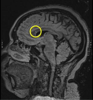
Figure 1: T2 FLAIR hyperintense lesion at the genu of the corpus callosum.
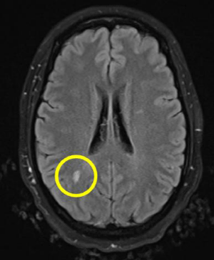
    Figure 2: T2 FLAIR hyperintense white matter lesion (9mm) in the right
subcortical white matter of the parietal lobe.
Discussion
Despite being the most common immune-mediated demyelinating disease of the central nervous system, MS is poorly characterized due to its heterogenous clinical and pathological features. MS often presents in young adults, affecting more women than men (2:1), with a clinically isolated neurologic syndrome, such as optic neuritis, numbness/paresthesia/weakness, or a brainstem syndrome such as internuclear ophthalmoplegia. These patients commonly present with a progressive or relapsing-remitting course of symptoms followed by partial or complete resolution. Typical symptoms include sensory disturbance in the extremities or unilateral face, visual deficits, bladder or bowel dysfunction, ambulatory difficulties due to motor weakness or gait disturbances, vertigo, pain and heat sensitivity. Depression or mood disturbance is also a common symptom in patients with MS.
The evaluation of MS should begin with a history and physical exam. Patients with suspected MS should undergo a brain MRI with gadolinium [1]. Positive MRI typically demonstrates focal white matter lesions, particularly in the periventricular and juxtacortical regions, corpus callosum and cervical spine. A diagnosis of MS can be made using the McDonald criteria which accounts for the number of clinical attacks coupled with the number and location of brain lesions observed via MRI of the brain and cervical spine [2]. If imaging and exam findings are inconclusive, lumbar puncture and CSF analysis may demonstrate MS specific oligoclonal bands which may assist the diagnosis [3,4]. Recent studies have also suggested the utility of testing visual evoked responses, optical coherence tomography, and autoantibody testing (AQP4 and MOG-IgG antibodies) in establishing a diagnosis of MS [4].
The differential diagnosis for a young patient who presents with weakness, pain and sensory disturbances includes infectious etiologies such as Lyme disease, Ehrlichiosis, HIV or tertiary syphilis; metabolic abnormalities such as vitamin B12 deficiency; heavy-metal toxicity; vasculitis; other autoimmune or inflammatory conditions such as SLE, or polyarteritis nodosa; neuroanatomical abnormalities such as Chiari malformation or other compressive vascular malformations or spinal cord lesions; and granulomatous diseases such as sarcoidosis [5].
Treatment of MS aims to delay neurologic decline and the rate of relapse. Acute exacerbations of MS (neurologic symptoms lasting longer than 24 hours with a preceding 30+ day period of clinical stability) are often treated with high-dose oral or intravenous glucocorticoids such as methylprednisolone, often followed by a prednisone taper. In patients that are not able to tolerate high-dose glucocorticoids, corticotropin injection gel may be administered either intramuscularly or subcutaneously. Patients with a relapsing-remitting forms of MS are often treated with disease-modifying therapies, such as dimethyl fumarate- which our patient received, interferon-beta, glatiramer or biologic medications such as natalizumab or ocrelizumab based on disease progression, and potential side-effects. In patients with refractory disease, switching to an alternative option may be beneficial.
It is also important to adopt a multi-disciplinary approach to manage the symptoms of MS which should account for possible onset of cognitive deficits, visual disturbances, fatigue, mood disorder including depression, bowel and bladder dysfunction, ambulatory dysfunction, and sensory changes especially in patients who may have progressive disease.
Take-Away
This case demonstrates the need for a thorough and rational approach to the clinical history, physical exam, and lab and imaging work-up when a patient presents with inconsistent or atypical features of MS, especially when prior neuroimaging is nondiagnostic. In such patients, additional testing for CSF specific oligoclonal bands, visual evoked potentials, optical coherence tomography or autoantibodies can be used to support the diagnosis(3,4). Clinically, it is important to note worsening or accumulating neurologic dysfunction which may represent progressive MS, as well as to obtain new diagnostic imaging when a patient may present with a new isolated acute exacerbation to examine the development of new demyelinating CNS lesions.
References
- Rovira À, Swanton J, Tintoré M, Huerga E, Barkhof F, Filippi M, et al. A single, early magnetic resonance imaging study in the diagnosis of multiple sclerosis. Arch Neurol. 2009; 66:587–592.
- McDonald WI, Compston A, Edan G, Goodkin D, Hartung HP, Lublin FD, et al. Recommended diagnostic criteria for multiple sclerosis: Guidelines from the international panel on the diagnosis of multiple sclerosis. Ann Neurol. 2001; 50:121–127.
- Thompson EJ, Freedman MS. Cerebrospinal fluid analysis in the diagnosis of multiple sclerosis. Adv Neurol. 2006; 98:147–160.
- Thompson AJ, Banwell BL, Barkhof F, Carroll WM, Coetzee T, Comi G, et al. Diagnosis of multiple sclerosis: 2017 revisions of the McDonald criteria. Lancet Neurol. 2018; 17:162–173.
- Miller DH, Weinshenker BG, Filippi M, Banwell BL, Cohen JA, Freedman MS, et al. Differential diagnosis of suspected multiple sclerosis: A consensus approach. MultScler. 2008; 14:1157–1174.