
Abstract
Dysplasia epiphysealis hemimelica (DEH) is developmental disorder characterized by osteocartilaginous over- gross in one or more epiphysis. The disease usually involves a single limb or is hemimelica (lateral or medial compartment) and lower extremities are more frequently affected than upper extremities. Here we present clinical and radiological findings for a male DEH patient at 12 year of age. The radiographs obtained at first presentation showed small osseous intra articular over growth at the base of the of the right knee, radiographs obtained at the second representation showed two osteocartilaginous masses at the tibial epiphysis. Two months after diagnosis the patient had surgery on his right knee to relieve the right knee pain and swelling, the histopathological diagnosis was consistent with osteocartilaginous. This case report presents imaging features and age related progression of DEH in this patient.
Keywords: Dysplasia epiphysealis Hemimelica; Trevor-fairbank; Radiography; Magnetic resonance imaging
Introduction
Dysplasia epiphysealis hemimelica (DEH) is also known as Trevor Fairbank disease [1,2], is a rare developmental skeletal dysplasia characterized by asymmetric osteochondoral over growth of epiphyseal cartilage DEH has an approximate incidence of one in one million and predominates in male (3:1) male: female). DEH can affect either a single joint or multiple joints the involvement is usually in a single limb or hemimelica lateral or medial compartment.
Radiographically, DEH is characterized by asymmetrical overgrowth of epiphyses with irregular ossifications.
Imaging studies play an important role in DEH diagnosis due to its characteristic appearance on radiographs. This case report presents the imaging features and age related progression of a male DEH patient diagnosed at 12 years of age.
Also the report highlights that, although DEH diagnosis is usually straight forward in terms of having atypical appearance, location, and distribution, the findings in the early stages can be subtle.
Case Presentation
This is 12 years old boy who presented to our hospital orthopedic out patient with his father referred from our primary care physician seeking medical advice and possible treatment options.
The father noticed a problem in his son’s knee has been raised to him by his son himself. This problem started when the boy noticed painless swelling (lump)on medial aspect of the right knee at the level of the joint before about 6 months which increasing in size and started to be painful with sport activity .
At presentation the boy was a bit shy but cooperative and holding good conversation and expressed his worries with assistance of his father.
On examination there are no associated congenital anomalies and no similar problem elsewhere in the body, only localized lump on the medial aspect of the right knee, hard in consistence, not mobile, not tender, and not pulsatile. He has painless full range of motion of the right knee. Stable ligaments of the right knee, no Neurovascular abnormality have been detected distally. Both limbs having the same length (Figure 1, 2).
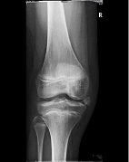
    Figure 1: Radiograph obtained when the patient was at 9 years at the first
OPD showed Osseous overgrowth at the medial tibial epiphysis at the right
knee joint.
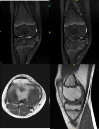
    Figure 2: Coronal (A) axial (B) coronal (C) sagittal (D) images demonstrate
osteocartilaginous masses at the right proximal tibia and distal femur
epiphyses.
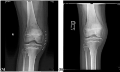
    Figure 3: Radiograph A 2 days after the operation, radiograph B about 1
month after the operation.
Detailed radiological report in radiology report.
Findings
First by x ray, it shows small osseous lesion at the medial side of the knee joint.
X ray followed by knee MRI with multiple sequences (axial PD FS, coronal T2 FS, sag T1, axial T2, axial T1, sag PD FS, coronal PD, axial FS, GR)which shows, multiple epiphyseal cortical irregularity of the femoral medial condyle affecting the posterior part, some are showing thin undermining low SI line please see (Figure 2d), and covered by hyaline articular cartilage, the same medial side of the knee joint , there are two osseous lesions medial and superior to the medial tibial plateau showing central signal follow the bone marrow and low SI cortex covered by thin layer following the hyaline articular cartilage on all pulse sequences, it displaces the medial meniscus superiorly and those lesions are located lateral to the medial collateral ligaments.
After necessary work up patient was taken to OR and open excision was done for 2 big pieces bony ones they were extra capsular pointing from the epiphysis covered with cartilage cap .the medial meniscus was paper film thin and elongated on the medial aspect of the femoral condyle. So after excision of exocytosis the service has been smoothening. And meniscus has been sutured back and stabilized in its position on medial tibial plateau. Then wound closed in layers and dressing has been applied.
Patient has been seen next day in the ward and sent home after that in a good condition.
Post-operative course was unremarkable clinically and radiologicaly, after one and half year the father was contacted and indicates his son in a good condition and a symptomatic.
Discussion
Dysplasia epiphysealis hemimelica which is also called trevor fair bank disease was first reported in 1926 by Monchet and Belot. Who described the condition as tarsomegalie based on its location, in 1950 Trevor reported 8 cases of the disease and named them taso–epiphyseal achalasis. In 1956 fair bank used the term dysplasia hemimelica (DEH) to describe this skeletal disorder because the disease also involves other joints and only half rather than the entire epiphysis.
DEH results in asymmetric osteocartilaginous overgrowth of the epiphysis and presents as skeletal deformity, pain, and restricted joint movement. DEH is considered to be a non-hereditary disorder, although the etiology of DEH is unclear, it may be associated with a congenital defect that affects the early stages of limb development during the fetal period or while chondrocytes proliferates without regulation, malignant transformation of DEH has not been reported.
References