
Abstract
Congenital radioulnar synostosis is a malformation that can limit the ability to perform daily activities. The condition involves the fusion of the proximal aspect of the ulna and radial head. The condition may result as a congenital defect or posttraumatic complication. Depending on the severity of the anomaly, treatment options include surgical intervention or conservative therapy. This case report discusses the initial presentation of congenital radioulnar synostosis in a 21-month old male. Imaging supports incomplete synostosis of the left forearm and the early stages in the right forearm.
Keywords: Congenital malformation; Radioulnar synostosis; Derotational osteotomy
Case Presentation
A 21-month old Asian male was referred to an orthopedic surgeon by his pediatrician for left arm pain. His parents noticed certain limitations in movements of the left arm about three weeks ago, specifically in tasks requiring supination such as eating finger foods. The father stated that when trying to gently assist in these movements, the patient would cry. The pediatrician referred the toddler after a limited range of motion exam and abnormal radiographic imaging. The family stated that they have been regularly taking the infant for checkups and no abnormalities were noted in the past. The parents denied any trauma to the left arm.
Review of systems was negative for: fevers, abnormal weight change, changes in appetite, changes in sleep patterns, changes in behavior, excessive fussiness, vomiting, diarrhea, other joint pains or signs of inflammation. There was no known family history of bone deformity or skeletal disease. Birth history included spontaneous vaginal delivery at 39 weeks with no complications. No significant past medical history was given. Surgical history was positive for circumcision. The toddler lived with both of his parents and did not attend daycare. There was no history of smoking or drinking in the household.
The toddler’s vitals were unremarkable. He appeared well nourished with a height and weight appropriate for his age group. On exam, the toddler was resting comfortablyon his father’s lap and appeared in no acute distress. He was awake, alert, and oriented. Skin exam was normal with no erythema, edema, or ecchymosis. There was no tenderness to palpation of the left or right elbow including the lateral epicondyle, forearm, and wrist. Passive range of motion of the left arm showed flexion and extension from 20 to 140 degrees, 80 degrees of pronation, and 0 degrees of supination. The right arm showedflexion and extension from 0 to 140 degrees, 50 degrees of pronation, and 30 degrees of supination. Musculoskeletal range of motion exam was completed without pain or crepitus. Bilateral motor exam was unremarkable with sensation to light touch in medial, radial, ulnar, and axillary nerve distributions. Active range of motion and strength could not be tested due to the age of the toddler.
Heart was regular rate and rhythm with no murmurs, rubs, or gallops. Lungs were clear to auscultation bilaterally with no rhonchi or wheezing. No clubbing or cyanosis with a capillary refill less than 2 seconds. Radiographic imaging was ordered in the office of the forearm bilaterally. The left forearm showed a proximal fusion of the radius and the ulna (Figure 1). The right forearm showed early stages of synostosis in the same area as the left (Figure 2).
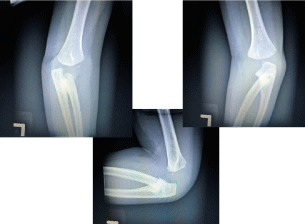
    Figure 1:  Shows the left AP, oblique, and lateral views of the left forearm.
The synostosis is apparent between the proximal ulna and the radial head.
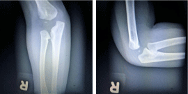
    Figure 2:  Shows the right AP and lateral views of the right forearm. Early
formation of the synostosis is apparent at the proximal ends of the radius
and the ulna.
Treatment of radioulnar synostosis consists of conservative management or surgical fixation (Figure 3). There are several factors when considering surgical treatment including age of the patient, severity of the malformation, and the functional limitation. Depending on the degree of the anomaly, conservative treatment with therapy may be appropriate. In this particular case, the family decided to delay any surgical intervention. Due to the young age of patient, surgery may not be the best option since the bone has not fully developed. The degree of malformation cannot be properly assessed due to his young age. It was recommended to postpone the surgery until the malformation has matured. The borders of the defect needed to be clearly visualized on radiographic imaging in order to prevent reoccurrence [1-6].
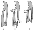
    Figure 3:  Shows the image depicted in Hung’s study of derotational
osteotomy. The procedure includes an osteotomy at the proximal radial
aspect and the distal ulna. Wires are then inserted through both the radial
and uln arstyloid, the bones are then reduced to a neutral position [2].
The limitation due to the anomaly also cannot be evaluated due to the young age of the toddler and inability to accurately test active range of motion. Surgical intervention is optimal during early childhood prior to school age in order to avoid any debilitation. Hung’s study evaluating derotational osteotomy found that, “The preoperative forearm rotation ranged from 65 degrees to 85 degrees pronation. The postoperative forearm rotation angle was corrected from 0 degrees to 30 degrees; the best end position appears to be 70 to 100% of pronation [2]”. The family was referred to a pediatric orthopedic surgeon to follow the infant’s development with serial plain radiographs and possible computed tomography scan.
Discussion/Conclusion
Radioulnar synostosis is a skeletal malformation resulting in the fusion of the proximal aspect of the ulna and radial head. The etiology of this condition can be post-traumatic or congenital. Post-traumatic radioulnar synostosis is most commonly as a result of a forearm fracture [6]. In this case report, the rare congenital anomaly resulted in limitation of movements, specifically pronation and supination. During the 37th week of embryonic development, there is a period of time prior to segmentation in which the radius and the ulna share a perichondrium [6]. Dysfunction in this sequence of events may cause failure in separation of the two bones followed by the ossification of the fusion.
There are multiple classifications of types of radioulnar synostosis depending on etiology. Wilkie defined two types of congenital radioulnar synostosis: type one refers to complete fusion of the proximal aspects of the radius and the ulna and type two refers to a partial union of bones as well as dislocation of the radial head [6]. The more commonly used classification of congenital radioulnar synostosis is defined by Cleary and Omer. The classification discussed by Siemianowicz et al.:
In typeI, there is a decreased size of the radial bone, the fusion does not involve bones; in type II there is a radioulnar synostosis, and the remaining bone structures do not reveal any other changes. In type III, there is a radioulnar synostosis, hypoplastic head of the radial bone, and posterior subluxation of the radial head. In type IV, there is a short radioulnar synostosis, mushroom-shaped malformation of the head of the radial bone, and anterior subluxation of the radial head [4].
There are two main methods of treatment for this condition: conservative or surgical. Conservative therapy may be beneficial in cases with minimal restrictions due to the malformation. A study evaluating the effect of surgical intervention on 34 patients with congenital radioulnar synostosis with an average age of 6 years and 3 months found that 78.8% of the participants had improved mobility after the procedure [2]. Hung’s study found that surgical intervention was beneficial when the forearm was fixed in pronation over 60 degrees and daily activities cannot be independently performed [2]. The recommended age for surgical intervention ranges from 3 to 10 years old depending on the surgeon but ideally before adolescent years to avoid complications. Complications of delayed surgical intervention include decreased functionality of forearm in activities of daily living as the child has learned how to compensate. Surgical intervention before the maturation of the synostosis may result in suboptimal surgical outcomes, decreased range of motion, and an increased rate of reoccurrence. Outcomes of patients who underwent the surgery older than six years old had a larger degree of pronation postoperatively [2].
When evaluating a patient with radioulnar synostosis, whether the etiology is post-traumatic or congenital, it is important to consider both treatment pathways. If surgical intervention is the preferred option, it is important to properly evaluate the degree of the malformation before proceeding. The fusion should be evaluated for distinct margins on imaging prior to undergoing a derotational osteotomy. Although congenital radioulnar synostosis is a rare malformation, if treated properly with surgical fixation and therapy the functional limitation associated with this condition can be reduced.
References
- Congenital radio-ulnar synostosis. National Center for Advancing Translational Sciences: Genetic and Rare Diseases Information Center. [Internet]. 2012 [cited 2 June 2016].
- Hung N. Derotational osteotomy of the proximal radius and the distal ulna for congenital radioulnar synostosis. J Child Orthop. 2008; 2: 481-489.
- McKean J. Taylor B. Radioulnar Synostosis - Trauma - Orthobullets.com [Internet]. Orthobullets.com. 2016 [cited 1 June 2016].
- Siemianowicz A, Wawrzynek W, Besler K. Congenital Radioulnar Synostosis, a case report. Polish Journal of Radiology. 2010; 75: 51-54.
- Spritz R. Familial radioulnar synostosis. Journal of Medical Genetics. 1978; 15: 160-162.
- Wurapa R. Radioulnar Synostosis: Background, Pathophysiology, Etiology [Internet]. Emedicine.medscape.com. 2016 [cited 1 June 2016].