
Abstract
A 63-year-old female presents after a palpable bulge along her bicep following a fall during an ice storm. She reported pain specifically with elbow flexion and supination of the wrist concerning a bicep injury. Given the palpable deformity, an ultrasound was obtained as there was concern for bicep tendon rupture resulting in retraction of the tendon into the mid-humerus region. However, it demonstrated intramuscular calcifications concern for myositis ossificans of the bicep. Further investigation with magnetic resonance imaging confirmed this conclusion. It was confirmed by ultrasound 18 months later. The age, gender, and mechanism of injury sustained by our patient differs from the normal course of myositis ossificans. Myositis ossificans is typically an athletic injury of a young male athlete located in the lower extremity. A typical presentation is related to trauma with severe contusion to a muscle as might be seen during a tackle in football. However, in our case her injury more so relates to a severe contusion from a fall as we might see in basketball when coming down from a rebound or football when making a catch.
Keywords: Myositis ossificans; Biceps; Muscle trauma; Contusion
Introduction
Myositis ossificans (MO) is a disease typically seen in young athletes injured during competition [1-5]. Most cases are seen in 15–40-year-old males with a high percentage being due to football injuries [2,3]. This disease process was first mentioned in the literature as early as 1905 when Jones and Morgan identified a benign ossifying tumor following a trauma [6]. Eight years later, three more cases of MO following trauma were documented [7]. In 1923, MO was classified into three forms: traumatic, nontraumatic, and neurotic [8]. Today the classification we know today includes myositis (fibrous) ossificans progressive, traumatica MO, MO circumscripta without history of trauma [9]. Traumatica MO is the most common subtype [1]. Non traumatica MO is typically found with underlying or antecedent illness such as a viral/bacterial infection or spasticity due to trauma related to cerebral palsy [8]. An atypical presentation or nonspecific imaging findings are more concerning for malignancy [1,10].
This benign condition results from severe contusion to muscle groups which can be specific to certain sports like the adductor muscles in horseback riders (Rider’s bone) and the deltoid for shooters (Shooter’s bone) [1]. The most common areas, however, are located in the lower limbs, thigh, and hip compartment due to high risk of injury at these locations [7,8] of these, the anterior thigh is the most common location [6]. MO is commonly seen in the skeletal muscle but can be seen in the tendons and subcutaneous fat. The pathophysiology of MO is incompletely understood, but there are two predominant theories. One predicts it occurs from an inappropriate differentiation of mesenchymal stem cells into chondrocytes and osteoblasts in an inflammatory rich environment following injury [1]. Another theory is that bone formation is most prominent at the periphery and margins of the lesion where they are rimmed by a monolayer of osteoblasts [8]. Typical initial presentation is pain and stiffness of the muscle and associated joint following a blunt tissue trauma such as a football player who gets hit directly in the thigh during a tackle [1,11,12], which may be diagnosed for a strain or contusion. Persistent pain longer than expected can alert the clinician that MO is present [1]. Less commonly associated weakness, numbness, lymphedema, and venous thromboembolic disease can occur when the lesion compresses nearby neurovascular structures. Symptoms tend to improve once the lesion becomes mature. Patients who present later in the lesion development may even be asymptomatic [1].
Case Presentation
A 63-year-old right hand dominant female sustained an injury to her right upper arm after falling during an ice storm initially experiencing pain in the right bicep and ipsilateral shoulder. She noticed a soft tissue bulge emerged three to four months later but did not seek care until one year after her injury. At that time, she reported point tenderness in the area overlying the anterior aspect of the upper arm where the bulge presented, mostly localized to the biceps area. She denied changes in strength but had difficulty lifting heavier objects as weeks passed due to pain. No overlying erythema, warmth, changes in ROM, or sensation changes were noted. The exam revealed a unilateral muscle bulge present over the right bicep. The strength of her right shoulder was normal but there was decreased strength with supination and elbow flexion. Dynamic testing of the shoulder revealed a positive Speed’s test (assessing for biceps tear/tendonitis) and Yergason’s test (assessing for biceps tear/tendonitis) [8]. Distal biceps tendon was able to be hooked. Out of concern for proximal biceps tear and palpable deformity, ultrasound demonstrated an intramuscular mass 2.0 x 1.1 x 0.9 cm in size (Figure 1). Follow up Magnetic Resonance Imaging (MRI) with IV contrast showed a 2.5 cm mild edema signal within biceps muscle around the hypointense center (Figure 2). A follow-up ultrasound was planned six months later, which demonstrated a mid humerus hyperechoic 3.1 x 3.2 x 1.2 cm mass with faint posterior acoustic shadowing suggesting internal calcification consistent with myositis ossificans (Figure 3). Treatment began with formal physical therapy, and subsequently following with repeat imaging to ensure resolution of calcification. Subsequent computerized tomography (CT) with IV contrast resulted in apparent chronic tear of biceps muscle belly with associated fibrosis without soft tissue calcification indicating resolution of MO. She received a subacromial steroid injection and referred to orthopedic surgery for surgical correction of the bicipital tear but was then lost to follow-up.
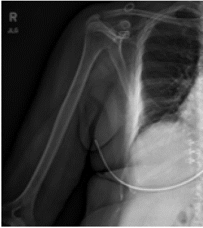
Figure 1: Xray.
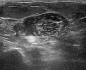
Figure 2: Ultrasound.
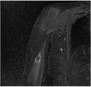
Figure 3: MRI.
Discussion
As with most musculoskeletal conditions, a systematic approach in regard to history, physical exam, and imaging are essential when diagnosing a shoulder injury. A history of a severe contusion occurs before changes are evident [6,13]. One looks for a classic triad of pain in affected muscle, palpable mass resulting from the presence of calcifications [8], and flexion contracture for MO (b). Ultrasound sometimes demonstrates a "checkerboard pattern" in an early lesion (Figure 2) [8,13]. The gold standard in mature stage and can be diagnostic on CT with presence of peripheral curvilinear calcific rim and central lucence resulting in an eggshell appearance which can be seen on MRI as well (Figure 3) [8,13] Initial calcification typically is visible on imaging within 2-6 weeks and becomes well-circumscribed by 2 months [13]. Radiographs or ultrasound can be repeated after 2-6 months to show pathognomonic circumferential calcification with lucent center and radiolucent cleft (string sign) is separate from the cortex of the adjacent bone. These imaging modalities can ultimately define resolution [13]. MRI can be beneficial as well if there is a concern for soft tissue pathology as source for pain such as concern for biceps rupture but typically will see a rim enhancement within first 3 weeks. Most cases of persistent lesions were diagnosed by a biopsy and not by imaging alone [8].
The initial history in our case regarding mechanism of injury, location of pain and initial symptoms is more congruent with biceps tendon rupture, biceps strain, rotator cuff injury, or intramuscular hematoma which are often seen after a trauma to the area. The palpable abnormality within the biceps muscle belly was the key to diagnosis and this led to the ultrasound. The differential can be divided into malignant pathologies including lymphoma, osteosarcoma, rhabdomyosarcoma and benign pathologies include calcified fibromatosis, local infections, biceps tendon tear, myositis ossificans, posttraumatic fibrosis, chronic intramuscular hematoma [14]. The most frequent clinical misdiagnosis is a tumor, especially extra-skeletal osteosarcoma which has similar clinical and pathologic characteristics [13]. Biopsy is indicated if there is persistence of palpable abnormality or imaging is not definitive. Histology displays a zonal pattern with periphery of lesion having mature trabeculae of lamellar and woven bone, and center demonstrating irregular mass of immature fibroblasts but no cellular atypia [8]. Of note, histological differentiation is the most difficult to separate from malignancy in the acute phase which is why traditional monitoring by imaging is initially recommended [6].
Congruent with many musculoskeletal conditions, NSAIDs are recommended for treatment in early stages. Physical therapy is recommended to be avoided in the very early stages as it can result in exacerbation of symptoms [8]. Despite this, often these cases are referred to physical therapy as treatment for an alternative suspected diagnosis such as in a muscular strain. The medical literature does recognize that assistive range of motion with pain-free arc of motion may begin as early as 48 to 72 hours [1]. Once the lesion matures, surgical excision can be recommended after at least six months, but these cases are typically reserved for symptomatic lesions that have failed nonsurgical treatment [1,8]. Surgical excision can be contemplated for intractable pain, compression of the important neurovascular structures, or decreased range of motion [1,8] local recurrence can occur in six to twelve months from the injury [1,8,14]. The prognosis of MO is related to size and location. Smaller lesions and those in the upper extremely are more likely to resorb completely whereas large lesions and those near insertion/origin of muscle are more likely to persist [2,6,15]. The condition is typically self-limited with an average disability length of MO in the anterior thigh is 73 days [15], but nonsurgical cases persist nearly two months [6].
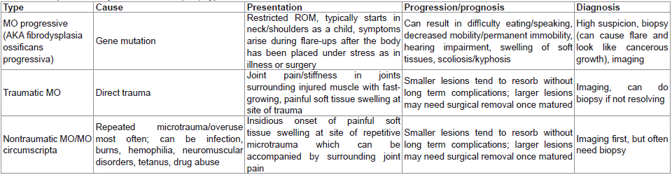
Table 1: Comparison of Myositis Ossificans (MO) Types.
Conclusions
Musculoskeletal injuries often occur from trauma and most injuries are usually self-limiting. However, if there is a persistence of pain, obvious deformity such as a palpable nodule or joint contracture, then further imaging is necessary. Characteristic findings of heterotopic bone formation on imaging indicate further investigation keeping myositis ossificans in mind. Not all cases occur in young athletes from competitive muscle trauma injuries. It must be contemplated in injuries of unusual mechanisms in uncommon locations, and for different age groups. Close observation clinically and radiologically is essential for all patients with concern of myositis ossificans.
References
- Walczak BE, Johnson CN, Howe BM. Myositis Ossificans. Jam Acad Orthop Surg. 2015; 23: 612-622.
- Folpe AL, Gown AM. Cartilaginous and osseous soft tissue tumors, in Goldblum JR, Folpe AL, Weiss WS, eds: Enzinger & Weiss’s Soft Tissue Tumors, ed 6. Philadelphia, PA, Elsevier. 2014; 917–946.
- Beiner JM, Jokl P. Muscle contusion injury and myositis ossificans traumatica. Clin Orthop Relat Res. 2002;: S110–S119.
- Huss CD, Puhl JJ. Myositis ossificans of the upper arm. The American Journal of Sports Medicine. 1980; 8: 419-424.
- Noble TP. Myositis ossificans, a clinical and radiological study. Surg Gynecol Obstet. 1924; 39: 795.
- Jones R, Morgan D. On osseous formations in muscle due to injury (traumatic myositis ossificans). Archive of the Roentgen. 1905; 9: 245.
- Coley WB. Myositis ossificans traumatica: A report of three cases demonstrating the difficulties of diagnosis from sarcoma. Ann Surg. 1913; 57: 305–337.
- Lewis D. Myositis ossificans. JAMA. 1923; 80: 1281–1287.
- Rehman N, Sadashiva H, Madakshira MG, Raman DK. Non-traumatic myositis ossificans. Autops Case Rep. 2021; 11: e2021316.
- Nuovo MA, Norman A, Chumas J, Ackerman LV. Myositis ossificans with atypical clinical, radiographic, or pathologic findings: A review of 23 cases. Skeletal Radiol. 1992; 21: 87–101.
- Arrington ED, Miller MD. Skeletal muscle injuries. Orthop Clin North Am. 1995; 26: 411-422.
- Edeiken J, Dalikna M, Karasick D. Edeiken’s Roentgen diagnosis of disease of bone. 4th ed. Vol Baltimore: Williams and Wilkins; 1990.
- Landolsi M, Mrad T. Traumatic myositis ossificans circumscripta (MOC). BMJ Case Rep. 2017; 2017: bcr2017219422.
- Ellerin BE, Helfet D, Parikh S, et al. Current therapy in the management of heterotopic ossification of the elbow: A review with case studies. Am J Phys Med Rehabil. 1999; 78: 259–271.
- Kransdorf MJ, Meis JM, Jelinek JS. Mysoitis ossificans: MR appearance with readiologic-pathologic correlation. Am J Roentgenol. 1991; 157: 1243-1248.