
Case Report
Austin ENT Open Access. 2025; 5(1): 1019.
Thyroid Ectopia of the Tongue: A Case Study and Scientific Analysis
Bekkali Z1,2*, Bencheikh R1,2, Boudinar H1,2, Elhafi Z1,2, Arkoubi Z1,2, Benbouzid MA1,2 and Essakalli L1,2
1Ent Head and Neck Surgery Department, Spécialities Hospital, Ibn Sina University Medical Center, Rabat, Morocco
2Faculty of Medicine and Pharmacy, Mohammed V University in Rabat, Morocco
*Corresponding author: Bekkali Z, Ent Head and Neck Surgery Department, Spécialities Hospital, Ibn Sina University Medical Center, Faculty of Medicine and Pharmacy, Mohammed V University in Rabat, Morocco Email: zineb.bekkali91@gmail.com
Received: April 06, 2025 Accepted: April 18, 2025 Published: April 23, 2025
Abstract
Lingual Thyroid Ectopia stands as a rare embryological anomaly, with an incidence ranging from 1 in 100,000 to 1 in 300,000. It is frequently linked with the absence of thyroid tissue in its conventional location, observed in around 70% of cases, and is notably more prevalent in females (3 to 7 females versus 1 male).
This particular case pertains to a 63-year-old female who has been experiencing swallowing discomfort for the past six months. Hormonal assessments revealed hypothyroidism (TSH at 8.326 μUI/ml), while cervical ultrasound suggested atrophic thyroiditis. A cervical CT scan confirmed thyroid atrophy and identified a lesion at the base of the tongue. The patient benefited from endoscopic reduction of the lesion, and subsequent histopathological examination definitively confirmed the thyroid origin of the resected mass.
Traditionally, lingual ectopia finds diagnosis in children, young adults, or around menopausal age. It typically presents as a mass at the base of the tongue, inducing local compressive signs often accompanied by hypothyroidism. Instances of hyperthyroidism and neoplastic involvement are, however, rare.
Thyroid scintigraphy using 99mTc or iodine-123 plays a pivotal role in diagnosis. Hormone therapy emerges as a viable strategy for achieving euthyroidism, impeding the growth of ectopic tissue, and circumventing the need for potentially intricate surgical excisions.
Introduction
Thyroid ectopia, a rare embryological anomaly stemming from the abnormal migration of thyroid cells, presents a diverse array of clinical manifestations. These range from congenital hypothyroidism, the leading preventable cause of intellectual disability, to asymptomatic cases diagnosed late in adult women. This complex pathology paints a varied picture, with basi-lingual localization notably prominent, representing 90% of ectopic cases.
Case Presentation
We report the case of a 63-year-old female with no notable medical history, presenting with a six-month history of dysphagia. Nasofibroscopy revealed a mass at the base of the tongue. Cervical ultrasound indicated an atrophic thyroid gland.
Cervical CT scan, performed with contrast enhancement, disclosed a localized lesion measuring 25x17x23 mm at the base of the tongue. This lesion was in proximity to the glosso-epiglottic fold and exhibited a macrocalcification. Cervicofacial MRI confirmed the presence of an oropharyngeal process centered on the base of the tongue, infiltrating the adjacent musculature and the right vallecula (Figure 1). Thyroid hormone assay corroborated hypothyroidism, with a TSH level of 8.326 μU/ml.
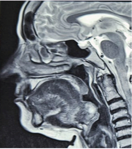
Figure 1: MRI image revealing an oropharyngeal process at the base of the tongue.
The patient benefited from endoscopic reduction of the lesion (Figure 2), and histopathological examination definitively confirmed the presence of thyroid tissue. The postoperative course was uncomplicated. Following the surgical reduction, the patient was placed under levothyroxine substitution therapy to ensure hormonal balance and to suppress TSH stimulation, thereby reducing the risk of regrowth of any residual ectopic tissue. Regular clinical and biochemical monitoring was scheduled, with TSH and free T4 assessments every three months. Cervical ultrasound was also planned at six-month intervals to monitor for any recurrence or growth of residual ectopic tissue. At the six-month follow-up, the patient remained clinically stable, euthyroid, and free of compressive symptoms.
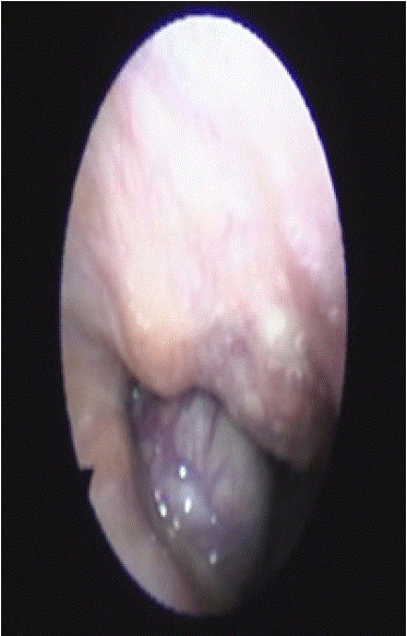
Figure 2: Endoscopic image of the lesion at the base of the tongue.
Discussion
After the second week of gestation, the thyroid gland emerges as an epithelial proliferation from the endodermal lining in the midline of the primitive pharyngeal floor, subsequently forming the "foramen cecum." It progressively migrates caudally and ventrally along the thyroglossal tract. By the seventh week, it reaches its final position below the thyroid cartilage, adjacent to the 5th and 6th tracheal rings [1,2].
Any disturbance in this migration can lead to thyroid ectopia [6]. Lingual thyroid is the most common, accounting for 90% of thyroid ectopias [1,5,7]. In the remaining 10% of cases, ectopia may be sublingual, submandibular, or, more rarely, mediastinal [1,3,6,7].
The pathogenesis of thyroid ectopia is still not fully understood [4]. Some suggest that maternal immunoglobulins targeting thyroid antigens may be responsible for this migration arrest [7]. Autopsy studies indicate a prevalence of thyroid ectopia ranging from 7 to 10% [1]. It is 3 to 4 times more common in females than males [1,2,4]. The average age of discovery is 40 years, with two peaks at 12 and 50 years [4,6].
Thyroid ectopia accounts for 75% of hypothyroidism cases in children [4]. Hypothyroidism secondary to physiological stress during puberty or illness may appear later in children with ectopic thyroid [5].
In most cases, symptoms of lingual thyroid occur during puberty, pregnancy, childbirth, or menopause [4,6]. The most frequent symptoms include oropharyngeal obstruction, dysphonia, dysphagia, or a sensation of a foreign body in the trachea and dyspnea [7]. Stridor may occur but is more common in the neonatal period [2].
Severe bleeding from an ectopic thyroid has also been reported, posing a potential life-threatening risk [4].
Imaging plays a crucial role in confirming the diagnosis, avoiding unnecessary biopsies [1,7]. It also guides the therapeutic strategy and detects possible malignant transformation [7].
Cervical ultrasound suggests the diagnosis based on the absence of the thyroid compartment and the presence of a mass overlying the base of the tongue with the same echogenicity as thyroid tissue [1]
Iodine-131 scintigraphy is the preferred method to confirm the diagnosis and determine the location of thyroid ectopia [1, 2]. It reveals basi-lingual hyperfixation with no fixation in the thyroid compartment. This method is sensitive and specific for the differential diagnosis between ectopic thyroid and other cervical masses [1].
In computed tomography, the mass appears discretely hyperdense due to its iodine richness. Intravenous contrast injection leads to intense enhancement of the mass [2]. MRI, with multiplanar acquisitions, allows better tissue and topographic characterization [1,2]. Lingual thyroid presents in MRI as a midline mass, well-defined with no signs of local-regional invasion, isointense in T1, hyperintense in T2, and enhances after gadolinium injection. Monitoring thyroid hormone and TSH levels is recommended in asymptomatic or small ectopia cases, especially since, in 70% of cases, this ectopia represents the only functional thyroid parenchyma. Surgical or radioactive iodine treatment can be considered in cases of persistent compression signs. In such cases, imaging shows an increase in mass size with a mass effect on neighboring structures and oropharyngeal lumen obstruction. Similarly, in cases of malignant transformation, MRI shows a heterogeneous aspect of the mass with signs of local-regional invasion.
Conclusion
The rarity of this embryological anomaly and its consequential local and functional implications underscore the importance of considering the diagnosis in the presence of hypothyroidism without palpable thyroid, even in adults. Suppressing hormone therapy proves effective in restoring euthyroidism, preventing the growth of ectopic tissue, often circumventing the need for sometimes intricate surgical excision.
In conclusion, long-term follow-up, whether under medical treatment or post-surgery, holds paramount significance in the comprehensive management of this condition.
References
- Cherif L, Lakhoua Y, Khiari K, Hadj-Ali I, Rajhi H, Kaffel N, et al. Thyroid ectopy: about two cases. Annales d’Endocrinologie. 2004; 65: 233–237.
- Ben Nasr M, El Ajmi W, Ben Amar F, Sellem A, Hammami H. Congenital hypothyroidism due to lingual thyroid ectopy: a case illustration. Médecine Nucléaire. 2022; 46: 99.
- Benzian Z, Benabadji N, Guittari H. Subhyoid ectopic thyroid with a normally located thyroid gland. Annales d’Endocrinologie. 2015; 76: 427.
- Oueslati S, Douira W, Charada L, Saïd W, Mlika N, Rezgui L, et al. Ectopic thyroid, Annales d’Otolaryngologie et de Chirurgie Cervico-faciale. 2006; 123: 195–198.
- Khessairi N, Oueslati I, Rached A, Sakka I, Rejeb O, Chihaoui M, et al. Thyroid ectopy: report of three cases. Annales d’Endocrinologie. 2017; 78: 337.
- Massine RE, Durning SJ, Koroscil TM. Lingual thyroid carcinoma: case report and review of the literature. Thyroid. 2001; 11: 1191–1196.
- Tadmori AE, Zermouni R, Ajdi F, Gaouzi A. Sublingual ectopic thyroid: case report of a young girl. Annales d’Endocrinologie. 2014; 75: 408.