
Research Article
J Drug Discov Develop and Deliv. 2023; 9(1): 1047.
A Novel Human Cell-Based Spheroid Model to Screen for Oncolytic Viruses and Oncolytic Viral Proteins
Nikita Narayan Naik¹; Nagaraj Nayak B²; Bharadwaja Vadloori³; Madhuri Subbiah²; Suresh Poosala¹; Akshay Sonawane³; Anjani Naru³
¹Acasta Health Pvt Ltd, Visakhapatnam, Andhra Pradesh, India
²National Institute of Animal Biotechnology, Hyderabad, Telangana, India
³Oncoseek Bio Pvt Ltd, Hyderabad, Telangana, India
*Corresponding author: Suresh Poosala Suresh Poosala , Acasta Health Pvt Ltd, Vyas Cancer Research Park, Plot No. 9&12 Layout vide TP No. 9/74,Survey No. 119, Krishna Nagar,Maharani Peta, Visakhapatnam, Andhra Pradesh, 530002, India. Tel: +91 9100851758 Email: suresh@acastahealth.com
Received: October 11, 2023 Accepted: November 20, 2023 Published: November 27, 2023
Abstract
Colorectal cancer is one of the major causes of cancer-related deaths worldwide and surgery has been the ideal treatment method. Oncolytic virus therapy uses a modified virus to specifically kill only cancer cells without harming the neighboring non-cancerous cells. A best example is Newcastle Disease Virus (NDV), an avian paramyxovirus which is seen as a promising oncolytic virus shown to combat many different types of cancers by in vitro and few in vivo animal studies. Traditionally, anticancer screening is done through in vitro cell line based assays in the lab and promising candidates are further tested in animal tumor models which is more expensive, time consuming and may not always reflect the truth due to species variations. Further results from the in vitro cell culture system may not always predict the outcome in animal models. 3D cell based models, i.e. spheroids are better than the cell lines as they can mimic the microenvironment of a tumor and can be used as an alternative to animal testing. In this study we demonstrated a novel human colorectal cancer spheroid model, which mimics the tumor microenvironment and better emulates the pathophysiology of human colorectal cancer, as a tool for identification of oncolytic agents. Through cytotoxic and colony forming assays we show the potential application of the spheroid model as a high throughput enabled anticancer screen.
Keywords: Oncolytic virus therapy; Cancer; Spheroid; NDV; Accessory viral protein
Introduction
Colorectal Cancer (CRC) is one of the major causes of cancer-related death worldwide in both men and women with a high incidence rate compared to other cancers. Colorectal, prostate, and lung cancer account for almost half (48%) of all cases in men. In the case of women, colorectal cancer, breast and lung cancer account for 51% of all new cases in the USA [1]. Colorectal cancer is the second-most common cause of cancer death in men and women combined in the USA. The 2023 Colorectal Cancer Statistics by the American Cancer Society summarized that although CRC incidences are overall declining, the number of CRC diagnoses at a younger age is rapidly increasing at a more advanced stage. The report also recommends accelerated progress against CRC by exploring the etiology of rising incidence in generations born since 1950. In addition, the recommendations include increasing access to high-quality screening and treatment among all populations [2].
The current main treatments for colorectal cancer are surgery and chemotherapy. However, these treatments have a poor prognosis as one-third of CRC patients have metastatic liver or lung tumors, reducing their overall survival rate, five years post-treatment, to only 50%. As noted by Song et.al, CRC, like many other malignancies, is a heterogeneous disease, which makes optimizing treatment against CRC and reducing related mortality a major challenge [3]. In recent decades, various types of immunotherapies have been gaining consideration, including oncolytic virus therapies to address the challenges mentioned [4,3].
The oncolytic virus kills only cancer cells without harming the neighboring non-cancerous cells. The main features of an oncolytic virus are that it selectively infects and replicates in tumor cells, and the virus further induces immunogenic death, necrosis, and autophagy; some oncolytic viruses also participate in apoptotic pathways. Newcastle Disease Virus (NDV), a highly infectious avian pathogen, is also a well-known oncolytic virus. NDV selectively infects and effectively kills tumor cells without affecting normal cells [3]. NDV has single-stranded RNA with a 15 KB genome. A mechanism of RNA editing by NDV effectively utilizes the compact genome. The P gene of NDV encodes 2 (accessory) nonstructural proteins, V protein, and W protein, via RNA editing. The V protein is known as a key virulence factor, in contrast very little is established about the function of NDV W protein function, the limited information available is from the studies on the Nipah virus W protein. A recent study by Yang et.al confirmed W protein expression in NDV and the unique subcellular localization of W proteins in virulent and avirulent NDV [5]. Further, the study by Nayak et.al, expressing W protein in NDV-infected DF1 cells, was the first to characterize the distinct features of W protein of moderately virulent NDV [6].
Traditionally, anticancer screening is done through in vitro cell line based assays in the lab and promising candidates are further tested in animal tumor models. However, these in vitro models are a trade-off between in vivo relevancy , cost and throughput. Due to the significant physiological differences between the in vitro, in vivo and clinical subjects, these in-vitro and in-vivo models often fail to replicate the human physiology faithfully leading to poor clinical transferability [7]. 3D cell based models, such as spheroids provide better in-vitro models compared to 2D in-vitro models as they can mimic the tumor microenvironment and can be used to refine the animal testing and eventually as an alternative.
In this study we demonstrated a novel human colorectal cancer spheroid model, which mimics the tumor microenvironment and better emulates the pathophysiology of human colorectal cancer, as a tool for identification of oncolytic agents. Through cytotoxic and colony forming assays we show the potential application of the spheroid model as a high throughput enabled anticancer screen. In addition, our study also addresses challenges, such as penetration into tumor bulk and adverse conditions in the tumor microenvironment, through novel spheroid models of human colorectal cancer. In the future, we intend to address other challenges like off-target effects and lack of specific predictive and therapeutic biomarkers [8]. Our study explores the oncolytic potential of NDV-K and the W protein through its apoptotic nature against colorectal cancer in the novel human colorectal cancer spheroid model post-infection and transient transfection.
Methods
Cell lines, Viruses, and Plasmids
HCT116 cells (procured from Atal Incubation Center, India) were revived, subcultured, and maintained in complete McCoy (Cat. No. AL0575, Himedia) media with 10% FBS, 2% Penstrep, and 1% Gentamicin in CO2 incubator at 37 degrees Celsius. Infection experiments were performed using Newcastle Disease Virus strain Komarov (NDV-K) (borrowed from Dr. Madhuri Subbiah, NIAB, Hyderabad). The transfection experiments were carried out with mammalian expression vector, pEYFP and pEYFP-W (W with N-terminal YFP tag) plasmids (borrowed from Dr. Madhuri Subbiah, NIAB, Hyderabad) using polyethylimine (PEI). The experiments were in triplicates and repeated twice.
Spheroids Formation
The cells were trypsinized at 70% confluency and 2000 cells (per well) were seeded in a 96-well ultra-low attachment plate (Cat. No. 174925, Thermo Fisher). The seeding of cells was followed by centrifugation of the plate at 1000 rpm for 5 minutes. After the centrifugation, the cells were observed for aggregation at the center by a phase contrast microscope. The seeded cells were incubated in a CO2 incubator at 37 degrees celsius for 72 hours.
PI Staining
Propidium Iodide (PI) was added at a concentration of 5μg/ml to the culture medium along with the cells while seeding for spheroid formation. So that the cells that make up the center of spheroids are exposed to PI. PI-exposed cells allow visualization of dead cells, as PI penetrates the plasma membrane of dying cells. PI (5μg/ml) was also added to growth media used to replenish 3D cultures after day 3 of spheroid seeding, ensuring that it was not limiting for the proliferating cells of the spheroids [9].
NDV Infection
The spheroids were infected with NDV-K on day 3 of spheroid seeding. Briefly, the spheroids were incubated with different MOIs (0 MOI, 10 MOI, and 20 MOI) NDV-K virus in basal McCoy media for 1 hour in a CO2 incubator at 37 degrees Celsius. The incubation was followed by media change, by removing 100 μl of the existing media and replacing it with 100 μl basal McCoy media. Imaging of the PI-stained, NDV-infected HCT116 spheroids was performed using a motic fluorescence microscope at 24 hrs and 48 hours post-infection. Furthermore, quantitative analysis of cell death of NDV-K infected spheroids (0 MOI, 1 MOI, 10 MOI, and 100 MOI) 72 hours post-infection, was performed by Cell titer glo assay and colony formation assay.
Co-Transfection and Transient Transfection
Transfection was performed using PEI (1 mg/ml) as a transfection reagent. For co-transfection, a complex of plasmid (pEYFP and pEYFP-W) and PEI was prepared with a ratio of 1:2 (0.75 μl and 1.5 μl respectively, for each well) in complete McCoy media (100 μl per well) and the complex was incubated at room temperature for 30 minutes, after thorough vortexing and short spin. The complex was mixed with the cells during seeding. The seeded cells were incubated in a CO2 incubator at 37 degrees celsius for the spheroid formation. Transient transfection was done on day 3 of spheroid seeding. The plasmid- PEI complex was prepared as mentioned earlier with a ratio of 1:2 (with 1 μl of plasmid and 2 μl of PEI, respectively, for each well) in basal McCoy media (100ul per well). This complex was added to the spheroids after the removal of 100 μl of the existing media. There were 4 sets of treatment, negative control (untransfected), mock transfected (only PEI), WYFP transfected (the complex of plasmid and PEI), and positive control (10 MOI NDV infection). Further, the W-transfected HCT116 spheroids without PI staining were subjected to colony formation assay for quantitative analysis.
Cell Titer glo Assay
NDV-K infected HCT116 spheroids were transferred onto a white bottom plate at 72 hours post-infection for the cell titer glo assay. The Cell titer glo assay (Cat. No. G9681, Promega) was performed as per the manufacturer's instructions. Briefly, the cells were lysed in the lysis buffer and the luminescence from the live cells was recorded after 30 minutes of incubation.
Colony Formation Assay
The protocol used for colony formation assay is a modified version of that used by Hu et.al. On day 5 of the spheroid seeding and 48 hours post-transient transfection, the spheroids were collected in sets (the replicates of each treatment together) in a 1.5 ml centrifuge tube. The collected spheroids were then washed with PBS, followed by accutase (1X, 200 μl per tube) treatment for 25 minutes and trypsin (50 μl 1X) treatment for 5 minutes at room temperature, to disperse the spheroids into a single cell suspension. Complete media (250 μl) with 10% FBS was added to the cell suspension to deactivate trypsin and the single-cell suspension thus formed was subjected to cell counting and 500 cells from each of the single-cell suspensions were seeded in a 6-well plate in triplicates for the formation of colonies. Media was changed after 4 days and the colonies formed were counted using a bright field microscope on day 8 of seeding [10].
Results
NDV Infection of HCT116 Spheroids
The Fluorescence microscopic images of PI-stained NDV-infected HCT116 spheroids (Figure 1) qualitatively showed a dose-dependent and time-dependent increase in the PI intensity, which represented the cell death in the spheroid. HCT116 showed significant susceptibility towards NDV infection starting from 1 MOI of NDV. Cell titer glo assay results of the NDV-infected HCT116 spheroids (Figure 2), showed a dose-dependent decrease in the percentage of viable cells.
![]()
    Time point

    P-Value


    24 HRS

    0.0013


    48 HRS

    0.0187


    72 HRS

    0.0048
Table 1: Statistical tests for the PI intensities (Mixed-design ANOVA test).
![]()
    Compared sets

    P-Value

    R square


    Untransfected &Mock

    0.0154

    0.8121


    Untransfected &WYFP

    0.0021

    0.9694


    Mock & WYFP

    0.0083

    0.9053
Table 2: Statistical tests for colony formation assay (Welch’s T-tests).
The colony formation assay from the single cell suspension of NDV-infected HCT116 spheroids (Figure 3) showed a dose-dependent decrease in the number of colonies formed. The results from the colony formation assay also corroborate with the Cell titre glo assay results of the NDV infected HCT116 spheroids.
W Transfection of HCT116 Spheroids
The untransfected spheroids are 5 days old and served as the negative control; Mock transfected are the spheroids transfected with only the transfection agent, PEI; WYFP transfected are spheroids with YFP plasmid with W protein and PEI; YFP transfected are spheroids with the YFP backbone plasmid and PEI; 10 MOI NDV infected spheroids served a positive control.
In Figure 4, there is an increase in cell death as represented by PI stain in WYPF transfected HCT116 spheroids compared to that in YFP transfected and Mock transfected and un-transfected HCT116 spheroids. The increase in cell death as represented by PI stain is time dependent in transfected HCT116 spheroids.
In Figure 5, there is an increase in cell death as represented by PI intensity represented graphically in WYPF transfected HCT116 spheroids compared to that in YFP transfected and Mock transfected and un-transfected HCT116 spheroids. The increase in cell death as represented by PI intensity is time-dependent in transfected HCT116 spheroids. In Figure 6, there is a significant difference in the number of colonies between; Untreated and Mock transfected, implying the cell death due to the transfection agent; Mock transfected and WYFP transfected, implying cell death due to the W protein. Based on the statistical analysis, the difference between Untransfected and WYFP; Mock and WYFP are significant as the p values are 0.0021 and 0.0083, which is less than 0.01 (99% confidence).
Discussion
The novel spheroid model of human colorectal cancer formed using the HCT116 cell line mimics the tumor microenvironment, thus addressing one of the challenges in Oncolytic virus therapy, adverse conditions in the tumor microenvironment. These cells would mimic the physiology of the colon and would facilitate the compactness of 3D spheroids playing an important role in matrix remodeling, thus facilitating a complex interplay between them. The spheroid will develop and thus perfectly recreate the pathophysiology of colon cancer. The novelty of the model lies in the exact emulation of the cell-cell interactions and the similar micro-environment of the in vivo situation concerning cell shape, adhesion, behavior, topology, and morphology, which other models, including animal models.
Our standardized protocol of transient transfection of the W protein for the spheroid model also addresses the challenge of penetration of the therapeutic agent into tumor bulk. The co-transfection along with the seeding of cells, ensures that the plasmid-PEI complex is exposed to the seeded cells that will make up the center of the spheroid. Since we are using transient transfection and the amount of plasmid with w protein is finite, as the cells divide the number of plasmids in each cell gets diluted. Thus, the second transient transfection after the formation of spheroid, on day 3 after seeding of the spheroid, ensures that the outer proliferation zone of the spheroid is adequately exposed to the plasmid-PEI complex. This protocol improved the efficacy of the transfection, introducing the plasmid with W protein in the cells, thus addressing one of the most common challenges of penetration of the therapeutic agent into tumor bulk.
The Fluorescence microscopic images of PI-stained NDV-infected HCT116 spheroids (Figure 1) qualitatively showed a dose-dependent and time-dependent increase in the PI intensity, which represented the cell death in the spheroid. The colony formation assay from the single-cell suspension of (strain Komarov) NDV-infected HCT116 spheroids (Figure 3) showed a dose-dependent decrease in the number of colonies formed. The results from the colony formation assay also corroborate with the Cell titer glo assay results of the NDV-infected HCT116 spheroids, Figure 2, which showed a dose-dependent decrease in the percentage of viable cells.
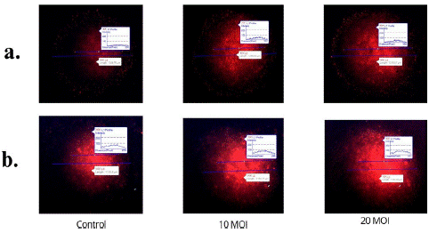
Figure 1: PI stained HCT116 spheroids at different time points (a: 24 hours, b: 48 hours) post NDV infection of different MOIs (0 MOI-Control, 10 MOI and 20 MOI).
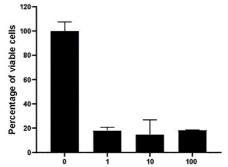
Figure 2: Cell titer glo assay of HCT116 spheroids 72 hours post NDV infection.
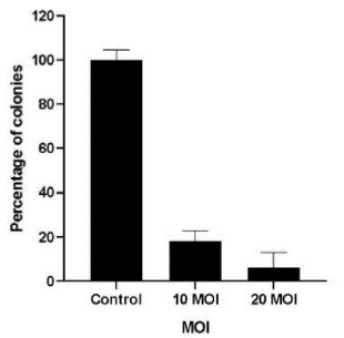
Figure 3: Colony formation assay from the single cell suspension of HCT116 spheroids 72 hours post NDV infection.
Fluorescence imaging of PI-stained W transfected HCT116 spheroids (Figure 4) at different time points (day 4, day 5, and day 6 respectively) post-transfection showed an increase in cell death, as represented by the increased intensity of PI stain and necrotic zone, in W transfected HCT116 spheroids compared to that in vector backbone transfected and Mock transfected and un-transfected HCT116 spheroids. The increase in cell death as represented by PI stain was also time- dependent in transfected HCT116 spheroids.
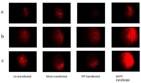
Figure 4: PI stained HCT116 spheroids at different time points (a: 24 hours, b: 48 hours, c: 72 hours) post W transfection.
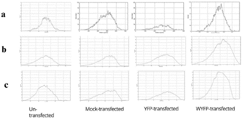
Figure 5: PI intensity of PI stained HCT116 spheroids at different time points (a: 24 hours, b: 48 hours, c: 72 hours) post W transfection.
The colony formation assay (Figure 6) seeded using the single cells suspension of the treated and control spheroids showed treatment-dependent significant differences in the number of colonies. The untransfected spheroids resulted in the highest number of spheroids. The number of colonies resulting from the mock-transfected spheroids was almost 25% less than that obtained from the untransfected spheroids, implying the cytotoxic effect of the transfection agent, PEI. The number of colonies resulting from the WYFP transfected spheroids was almost 30% less than that from the mock transfected and 50% less than that from the untransfected spheroids, implying the significant apoptotic nature of W protein and therefore suggesting the Oncolytic potential of W protein.
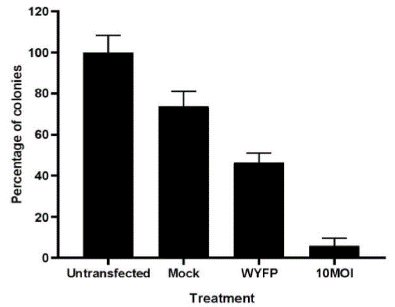
Figure 6: Colony formation assay from the single cell suspension of HCT116 spheroids 48 hours post transient transfection (day 5 spheroids).
Qualitative and quantitative analysis of cell death/viability of transfected spheroid showed significant cell death compared to controls, demonstrating the apoptotic nature of the W protein of NDV. Based on the statistical analysis, the difference between Untransfected and WYFP; Mock and WYFP are significant as the p values are 0.0021 and 0.0083, which is less than 0.01 (99% confidence).
To conclude, this study successfully standardized the amount and dosage of the NDV accessory viral protein resulting in significant cancer cell death in a novel human colorectal cancer spheroid model, which mimics the tumor microenvironment and better emulates the pathophysiology of human colorectal cancer. In the future, we will develop a recombinant oncolytic virus expressing the novel NDV accessory viral protein for further ex-vivo and in-vivo experiments with human-based and human-relevant data generation.
Author Statements
Acknowledgments
The authors would like to thank Dr. Madhuri Subbiah, NIAB, Hyderabad for lending us the plasmids and viruses. We would also like to thank Dr. Nagaraj Nayak for his contribution to the research work.
Declaration of Conflicting Interests
The author(s) declared no conflicts of interest with respect to the research, authorship, and/or publication of this article.
Funding
The author(s) received no financial support for the authorship, and/or publication of this article. The funding for the research was from self-investment.
References
- Siegel RL, Miller KD, Fuchs HE, Jemal A. Cancer statistics, 2022. CA Cancer J Clin. 2022; 72: 7-33.
- Siegel RL, Wagle NS, Cercek A, Smith RA, Jemal A. Colorectal cancer statistics, 2023. CA Cancer J Clin. 2023; 73: 233-54.
- Song H, Zhong LP, He J, Huang Y, Zhao YX. Application of Newcastle disease virus in the treatment of colorectal cancer. World J Clin Cases. 2019; 7: 2143-54.
- Geevarghese SK, Geller DA, de Haan HA, Hörer M, Knoll AE, Mescheder A, et al. Phase I/II study of oncolytic herpes simplex virus NV1020 in patients with extensively pretreated refractory colorectal cancer metastatic to the liver. Hum Gene Ther. 2010; 21: 1119-28.
- Yang Y, Xue J, Teng Q, Li X, Bu Y, Zhang G. Mechanisms and consequences of Newcastle disease virus W protein subcellular localization in the nucleus or mitochondria. J Virol. 2021; 95: e02087-20.
- Nayak BN, Rajagopal K, Shunmugasundaram R, Rao PL, Vaidyanathan S, Subbiah M. Molecular characterization suggests kinetic modulation of expression of accessory viral protein, W, in Newcastle disease virus infected DF1 cells. Virusdisease. 2023; 34: 236-47.
- Naik NN, Vadloori B, Poosala S, Srivastava P, Coecke S, Smith A, et al. Advances in animal models and cutting-edge research in alternatives: Proceedings of the Third International Conference on 3Rs Research and Progress, Vishakhapatnam, 2022. Proceedings of the third international conference on 3 Rs research and progress, Vishakhapatnam, 2022. Altern Lab Anim. 2023; 51: 263-88.
- Goradel NH, Baker AT, Arashkia A, Ebrahimi N, Ghorghanlu S, Negahdari B. Oncolytic virotherapy: challenges and solutions. Curr Probl Cancer. 2021; 45: 100639.
- Brüningk SC, Rivens I, Box C, Oelfke U, Ter Haar G. 3D tumour spheroids for the prediction of the effects of radiation and hyperthermia treatments. Sci Rep. 2020; 10: 1653.
- Hu L, Sun S, Wang T, Li Y, Jiang K, Lin G, et al. Oncolytic Newcastle disease virus triggers cell death of lung cancer spheroids and is enhanced by pharmacological inhibition of autophagy. Am J Cancer Res. 2015; 5: 3612-23.