
Case Report
Austin J Dent. 2025; 12(1): 1189.
Cervicofacial and Mediastinal and Pericardial Emphysema after Fail to Attempt Dental Implant Removal: A Case Report
Keosetha T, Sandeth P, Sophal Y, Sina C, Saroth T, Ping Bushara* and Pengleang C
University of Health Sciences, Cambodia
*Corresponding author: Ping Bushara, University of Health Sciences, Cambodia Email: busharaping@gmail.com
Received: July 25, 2025 Accepted: August 14, 2025 Published: August 18, 2025
Abstract
Although emphysema is a rare occurrence in dental practice, it is most commonly associated with surgical procedures involving the use of high-speed air-driven handpieces in which the air is forced into the mucoperiosteal flap. This can lead to the rapid development of soft tissue swelling at the surgical site, accompanied by crepitus characteristic on palpation. Entrapped air may then migrate along the cervicofacial connective tissue planes, extending into the pneumomediastinum, and occasionally involving the peri-visceral and chest wall areas, resulting in subcutaneous and profound emphysema.
Introduction: Surgical emphysema, or subcutaneous emphysema, occurs when air or gas becomes trapped in the subcutaneous tissue, often following surgical procedures in the oral and maxillofacial region. It can also arise spontaneously from self-induced actions, such as forceful nose blowing. Dental procedures involving airdriven instruments are a recognized cause of this condition, though it remains rare.
Case Report: In this report, we present a case with a rare, life-threatening complication. A 65-year-old woman presented with progressive facial and cervical swelling, pain, and dysphagia approximately 8 hours after a failed dental implant removal. Imaging confirmed extensive subcutaneous emphysema extending into the mediastinum. The patient was treated conservatively with supplemental oxygen, analgesics, and antibiotics under close monitoring. The patient recovered without further complications and was discharged on the ninth day.
Conclusion: Subcutaneous emphysema is characterized by rapid onset of soft tissue swelling with crepitus on palpation. While often self-limiting, severe cases may involve deeper anatomical structures, necessitating appropriate management. This case highlights the importance of early recognition and intervention to prevent potentially life-threatening complications. Surgeons must be aware of this risk and prepared to manage it effectively.
Keyword: Dental implant removal; Cervicofacial emphysema; Mediastinal emphysema
Introduction
Surgical emphysema, also known as subcutaneous emphysema, occurs when air or gas is trapped in the subcutaneous layer of the skin. This condition often arises from surgical procedures, trauma, or infections, particularly in the oral and maxillofacial region. However, it may also occur spontaneously from self-induced actions such as forceful nose blowing or playing wind instruments [1,2]. Surgical emphysema resulting from dental treatment has been associated with the use of high-speed air-driven handpiece instrument and/or air directed at the surgical site. Dental procedures most frequently associated with emphysema include tooth extraction, restorative dentistry, and endodontic treatment [3] increasing intra-oral pressure, such as blowing up a balloon, during the early post-operative period can lead to this complication [2]. A systematic review published in the British Dental Journal (2021), analyzing cases from 1993 to 2020, found that 54% of 135 reported cases of subcutaneous emphysema resulted from dental extractions, particularly surgical extractions. The majority of these cases were iatrogenic, with 51% attributed to the use of air-driven handpieces and 9% to air-syringes. Other factors, such as nose blowing, accounted for 10% of the cases [4].
This review also identified that air polishing or prophylaxis systems accounted for 3.7% of cases—half of which occurred during dental implant maintenance, while the remainder were related to routine periodontal treatments. The use of dental lasers with air-cooling systems contributed to 3% of cases [4].
Although mild subcutaneous emphysema can resolve spontaneously, this complication may spread to deeper fascial spaces leading to serious consequences if left unnoticed or misdiagnosed. In more severe cases, referral to oral and maxillofacial surgeon is advised for hospital admission and close monitoring.
Case Presentation
A 65-year-old female presented to the Oral and Maxillofacial Surgery (OMFS) department of Preah Ang Duong hospital with bilateral, progressive facial and cervical swelling with pain, dysphagia, and limited neck movement. The symptoms developed approximately 8 hours after undergoing an attempted dental implant removal. The patient denied dyspnea or hoarseness. The implant, located in the right anterior mandible and positioned lingually beyond the fixed porcelain prosthesis rim, had been surgically accessed under local anesthesia by the referral dentist.
A conventional dental air-driven handpiece was used to drill the prosthesis and peri-implant bone. The procedure encountered significant difficulties. After an hour of unsuccessful effort, the dentist was unable to remove the implant. The procedure was halted with plans to continue the following day.
On presentation, the patient was afebrile and not in respiratory distress. There was bilateral buccal and diffuse cervical swelling, with crepitus on palpation extending to the infratemporal, cheeks, neck, and anterior chest wall. No erythema was noted over these areas. Intraoral examination showed a normal mouth opening with a wound defect at the right floor of the mouth, suggesting an incomplete removal of the dental prosthesis and implant (Figure 1).
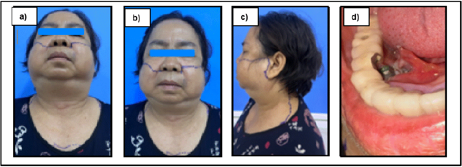
    Figure 1: Clinical photograph on arrival. A) through C) demonstrating severe bilateral cervicofacial swelling. D) Intraoral photograph presented with incomplete
removal of the mandibular prosthesis.
A head, neck and chest computed tomography (CT) scan was performed to assess the head, neck and thoracic region. The imaging revealed the extensive subcutaneous emphysema involving the bilateral temporal regions and deep cervical spaces, including the masticatory, parotid, parapharyngeal, retropharyngeal, and carotid spaces, with extension into the mediastinum and pericardium spaces, however there is no significant airway narrowing was noted (Figure 2).
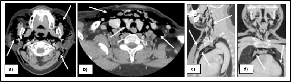
    Figure 2: Head, neck and chest CT scan presented severe emphysema involving the bilateral facial spaces and deep cervical spaces, including the masticatory,
parotid, parapharyngeal, retropharyngeal, and carotid spaces, with extension into the mediastinum and pericardium spaces.
The clinical impression was consistent with extensive surgical emphysema secondary to the surgical removal of dental implants. The patient was admitted for close airway monitoring. Management included the administration of supplemental oxygen and the initiation of intravenous analgesic and prophylactic antibiotics – specifically ceftriaxone and metronidazole. There was no progression of the emphysema, allowing for the continuation of conservative management (Figure 3).
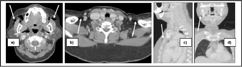
    Figure 3: A follow up CT scan on postoperative day seven was demonstrated significant improvement on subcutaneous emphysema, with minimal air was
remained.
On day seven, a follow-up CT scan was done. The subcutaneous emphysema was almost resolved dramatically, with only minimal residual air was remained. The retained implant fragment was performed under local anesthesia without complication. The patient continued to recover and was discharged on day nine with instructions for outpatient follow-up until complete clinical and radiographic resolution of the subcutaneous emphysema (Figure 4).
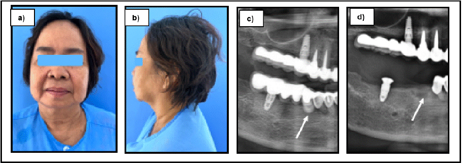
    Figure 4: a) and b) clinical extraoral photographs were obtained on postoperative day 7 follow-up, illustrating significant resolution of cervicofacial edema with
only slightly residual swelling. a) and b) pre and postoperative OPGs were demonstrated the removal of remain implant and prosthesis.
Discussion
Subcutaneous Emphysema is characterized by the rapid onset soft tissue swelling, typically appearing within seconds or minutes. The key clinical sign is crepitus or crackling on palpation, which differentiates it from other conditions like angioedema, anaphylaxis, or hematoma, where such a sign is absent. Discomfort levels may vary among patients
based on the extent of air infiltration. Although the air is typically reabsorbed over time from the surrounding tissues, however, serious cases may extend to deeper compartments such as the mediastinum – and, rarely, the pleural, pericardial, or peritoneal cavities – leading to complications like pneumomediastinum or airway compromise [3,5].
• Clinical examination: The differential diagnosis of emphysema includes conditions that cause sudden swelling such as anaphylaxis, angioedema, and internal hemorrhage. The specific context in which the swelling occurs is crucial for making an accurate diagnosis. Crepitus, characterized by a crackling sensation upon palpation, is the most critical distinguishing feature and is typically absent in cases of angioedema and hematoma formation.
• Investigation toward definite diagnosis:
• Imaging: Conventional X-rays, CT scans, or MRI may be used to assess the extent of air presence and rule out other conditions.
• Management strategies and literature comparison
o In this case, conservative management was chosen due to the patient's stable airway, absence of infection, and the self-limiting nature of the emphysema. This management is supported by several reports in the literature, which emphasize close monitoring, oxygen therapy to promote air resorption, and antibiotic coverage to prevent secondary infections.
o Ceftriaxone and metronidazole, a common prophylactic regimen in oral and maxillofacial infections, were selected for broad-spectrum coverage against both aerobic and anaerobic organisms.
o Literature showed that while most subcutaneous emphysema cases are mild, severe forms can rapidly progress and require hospitalization. Dental practitioners must be vigilant for signs and symptoms of these complications and be prompt to refer the patient in an effective manner. Misdiagnosis or delayed intervention can lead to life-threatening outcomes.
o Detail the management strategies employed in this case, including the use of supplemental oxygen, intravenous antibiotics, and close monitoring.
o Discuss why conservative management was chosen in this case and how it aligns with current best practices.
o Explore the rationale behind the choice of antibiotics (e.g., to prevent secondary infection due to the presence of air in soft tissues).
o Compare this case with other reported cases in the literature. Discuss the frequency of subcutaneous emphysema in dental procedures based on previous studies,
o Emphasize the importance of early recognition of subcutaneous emphysema by dental professionals.
o Discuss the potential for misdiagnosis and the consequences of delayed treatment, which could lead to life-threatening complications.
o Reinforce the need for prompt referral to specialists, such as oral and maxillofacial surgeons, when subcutaneous emphysema is suspected.
• Preventive measures
o Avoiding the use of high-pressure air in the surgical sites
o Avoiding the use of air-driven handpieces during surgery
o Ensuring proper technique during high-risk procedures
o Instructing the patient to avoid increasing intraoral pressure postoperatively (e.g., forceful nose blowing, balloon inflation, or playing wind instruments).
o Discuss preventive measures that can be taken during dental procedures to reduce the risk of subcutaneous emphysema. This might include careful handling of airdriven instruments, avoid excessive air pressure, and ensure proper technique during high-risk procedures.
o Mention the importance of patient education on avoiding actions that increase intraoral pressure (e.g., forceful nose blowing, playing wind instruments) in the postoperative period.
Conclusion
Understanding, early recognition, and prompt management of the subcutaneous emphysema are crucial in preventing life-threatening complications and ensuring patient safety.
Dental practitioners, who are uncertain about the management and potential complications, should promptly refer the case to oral and maxillofacial surgeons.
Surgeons must be vigilant this rare but potentially life-threatening complication and be prepared to ensure patient safety and positive outcomes.
References
- Shafto CE. Surgical emphysema of the neck and chest wall following dental extraction. Br Dent J. 1945; 78: 364–365.
- Shovelton DS. Surgical emphysema as a complication of dental operation. Br Dent J. 1957; 102: 125–129.
- Stanton DC, Balasanian E, Yepes JF. Subcutaneous cervicofacial emphysema and pneumo-mediastinum: a rare complication after a crown preparation. Gen Dent. 2005; 53: 122–124.
- Jones A, Stagnell S, Renton T, Aggarwal VR, Moore R. Causes of subcutaneous emphysema following dental procedures: a systematic review of cases 1993-2020. Br Dent J. 2021; 231: 493–500.
- Busuladzic A, Patry M, Fradet L, Turgeon V, Bussieres M. Cervicofacial and mediastinal emphysema following minor dental procedure: a case report and review of the literature. J Otolaryngol - head neck Surg = Le J d’oto-rhinolaryngologie Chir cervico-faciale. 2020; 49: 61.