
Case Report
Austin J Dent. 2025; 12(1): 1187.
Traumatic Pseudoaneurysm of Internal Maxillary Artery Following a Condylar Fracture: A Rare Case Report
Landge JS¹, Meshram AA¹*, Chauhan S², Shah KM¹ and Pedamkar K²
¹Department of Oral and Maxillofacial Surgery, Government dental college and hospital, Ghati medical campus, Chh, Sambhaji Nagar-431001, India
²Department of ENT, Grant Government Medical College, JJ Marg, Nagpada, Mumbai, India
*Corresponding author: Meshram AA, Department of Oral and Maxillofacial Surgery, Government Dental College and Hospital, Ghati Medical Campus, Chh. Sambhaji Nagar-431001, India Email: anjalimeshram013@gmail.com
Received: January 06, 2025; Accepted: January 28, 2025 Published: January 31, 2025
Abstract
Traumatic pseudoaneurysms in the head and neck regions are very rare. This case reports presents 17 yr old female who was diagnosed as having pseudoaneurysm of the internal maxillary artery. A pseudoaneurysm is characterized by a well-organized pulsatile mass that appears after a traumatic event. These lesions are frequently misdiagnosed as hematomas or abscesses. Audible bruit and pulses are indicative of an aneurysm. Such false aneurysm ruptures result in substantial morbidity. Management either embolization or surgical resection is the course of treatment. Endovascular embolization is recommended is indicated in deep seated lesion or lesions with high morbidity. Despite being an invasive process, surgical resection is thought to be an alternative to embolization.
Keywords: Pseudoaneurysm; Embolization; Internal Maxillary Artery
Introduction
The largest and terminal branch of the ECA is called the IMA. The proximal mandibular, middle pterygoid, and terminal pterygopalatine segments are its three main segments. It begins behind the mandible at the distal ECA bifurcation.5. The IMA splits into branches that supply the nose and deep face before terminating within the pterygopalatine fossa. The distal IMA is a major source of potential collateral blood flow from the external to the internal carotid artery via the inferolateral trunk and vidian artery. It also has numerous anastomoses with other ECA branches, such as the facial artery. Additionally, the distal IMA has anastomoses with the ophthalmic artery via the ethmoid artery.
"A focal, irreversible dilatation of an arterial wall is called an aneurysm." The tunica externa, media, and intima—the three parts of the artery wall—will all be present in a "true" aneurysm while a pseudoaneurysm may have one or two components [1-3].
The etiology may be (1) a blunt trauma to vessel, (2) iatrogenic transection of the vessel wall, (3) radiation injury and/or infection causing erosion of the vessel wall.
Common vessels involved are: (1) Facial artery, (2) superficial temporal artery, (3) descending palatine artery, (4) internal maxillary artery (first and last parts), and (5) internal carotid artery (ICA) [3,5,6].
Despite the great frequency of mandibular condylar fracture and the proximity of the maxillary artery to the fracture site, the occurrence of the false aneurysm to this artery is very rare [7].
Pathogenesis
The medial displacement of the fractured bone fragments can injure the internal maxillary artery or its major branches. The mandibular condylar region becomes the site of bleeding from the fractured bone ends and damaged/ruptured vessel until the pressures inside and outside the vessel equalize, which causes tissue tamponade. Aneurysmal sac are formed by cellular components during the reparative process of a ruptured vessel and a fibrous capsule is formed by the inflammation surrounding the hematoma the center of the clot liquefies. The lesion becomes pulsatile due to increased arterial pressure and “jetting” of blood through the sac, and auscultation detects a bruit. The formation of well-formed sac or pseudoaneurysm usually takes a long period, may be months or years after the trauma. fragments are displaced medial and anterior to right TMJ, angle of mandible and displaced comminuted fracture of para symphysis. The patient was treated for fracture of right para symphysis of the mandible by open reduction and internal fixation [2].
Patient was placed on intermaxillary fixation (IMF) for Angle fractures and condyle fracture. Patient had persistent swelling in front of the right ear since then. The patient reported to us for treatment of swelling on the right side of the face and also for the removal of IMF. Swelling was gradually increase in size, nontender and pulsatile in nature. CT- BRAIN angiography was advised to identify feeding vessels. CT-BRAIN angiography suggestive of heterogeneous lobulated lesion with hyperdense content of blood attenuation within, measuring 5x 3.8 x 5.4 cm (AP TRA CC) is seen involving the right masticator, parotid spaces and infratemporal fossa adjacent to right TMJ were performed findings are suggestive of post traumatic pseudoaneurysm formation with feeders from posterior auricular artery. The patient underwent additional imaging in the form of a carotid angiogram for detailed assessment of pseudoaneurysm which revealed an large pseudoaneurysm originating from internal maxillary artery.
Given the pseudoaneurysm's size and location along the trunk of the internal maxillary artery, the interventional radiologist team decided to proceed with coil-embolization.
Routine access through the femoral artery was obtained and a catheter was navigated to the common carotid artery. From this position, an angiogram was completed to survey details of size and location of aneurysm and the interventional radiologist team coiled the pseudoaneurysm for definitive management. Embolization was done through the microcatheters over microwire with 6 mm x 14 cm and 3 mm x 14 cm coils (Figure 2,3).
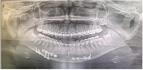
    Figure 1: Patient who reported with a complaint of increasing swelling in
front of the right ear since 1 month.
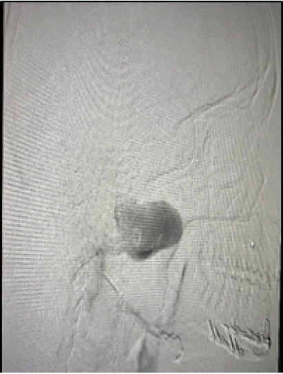
Figure 2: Before embolization.
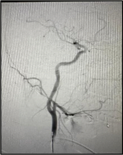
Figure 3: After embolization
Postprocedural angiogram reveals complete exclusion of the pseudo aneurysm.
Ater that patient is planned for excision of hematoma as well as also the open reduction and internal fixation for the ramus and angle fracture (Figure 4).
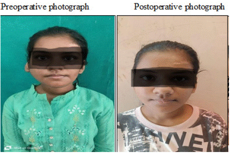
Figure 4: Preoperative photograph, Postoperative photograph.
Discussion
Posttraumatic aneurysms are all types of false aneurysms. Blunt force is usually the reason when there is not enough cushioning between the vessel and the bone since there is not much muscle tissue underneath [7]. It's also important to draw attention to high explosive military munitions, which rarely find usage in civilian settings and can result in a variety of horrendous injuries [8]. Along with idiopathic spontaneous aneurysms, reports of autoimmune diseases and subsequent infections have also been made [5,6]. It's crucial to remember that while pulsatile scalp masses and sudden-onset neck swelling, including significant or moderate bleeding, can be signs of posttraumatic pseudoaneurysms, they did not manifest in our case [8].
Dermoid or epidermoid cysts, eosinophilic granuloma, hematoma, abscess, aneurysm, arteriovenous fistula, encephalocele, lymphoid hyperplasia, and sinus pericrania should all be considered in the differential
Diagnosis [3,6,8]. MRI angiography, which demonstrates fusiform dilatation and turbulent intraluminal artery flow in non-thrombosed aneurysms, is one diagnostic method that can be used. Important details on the vessel of origin, luminal morphology, and relationship to surrounding osseous and soft tissue structures can be obtained by CT angiography [8]. Though it might be less helpful in situations with thrombosed aneurysms, digital subtraction angiography is thought to be the gold standard for characterizing these lesions and distinguishing them from arteriovenous malformations, which also manifest as pulsatile subcutaneous masses [7,8].
Hunter, who demonstrated the benefits of proximal arterial ligation in the 18th century, carried out the first attempts to treat pseudoaneurysms of the external carotid artery. Simple resection of the aneurysm, coil embolization [9], direct thrombin injection [10], proximal ligation of the parent artery, or aneurysm trapping are among the treatment options for traumatic pseudoaneurysms.
Particulate embolization is more frequently employed as a preoperative treatment to reduce the amount of peri- operative blood loss, while it can be utilized as a definitive operation as well. This method enables embolization of veins in close proximity to the site of bleeding. The material is intended to be deposited within the network of blood vessels. A catheter inserted into the femoral artery is used to do this. A contrast agent is used to locate the feeder channel while the catheter is progressed until it reaches the common carotid artery. The coils are conveyed via a microwire and a microcatheter. The formation of an embolus, around the coil stops the blood supply to the pseudoaneurysm. Embolization materials can be categorized as fluids that completely fill the lesion, particulate agents and mechanical devices. Few examples are gel foam, gelatin, Dacron fibers, N-butyl cyanoacrylate, autologous clot, polyvinyl alcohol, platinum complex coils, Guglielmi detachable coils, and more recently electronically detachable platinum micro coils [6,8].
Using small-sized particles is essential to preserve the parent and second-order arteries while achieving the most distal vascular blockage. When there is no visible flow into the artery's distal branches, the surgery is deemed to be finished.
Coil occlusion, rather than particulate embolization, would be the endovascular treatment of choice. A major risk with endovascular occlusion is movement of the occlusive material. Embolization is usually associated with local tissue infarction. Surgical excision at this site can be problematic although Weber et al. (1997) state that complete excision of a pseudoaneurysm with ligation of the proximal and distal parts of the artery is the recommended treatment.
In trained hands, morbidity associated with such treatment is almost negligible (0.03% mortality and 1.73% morbidity) and results are promising [2]. Common complications include injury to vessels, inadvertent embolization of ECA or ICA, dislodgement of coils or embolus, infarction, etc.
Conclusion
Proper monitoring and early detection. Therefore, appropriate decision-making through teamwork comprising surgical specialists and interventional radiologists is required for treatment planning of traumatic aneurysm of the internal maxillary artery. A carefully planned and executed access osteotomy will delineate the aneurysmal sac in its entirety.
References
- Zachariades N, Rallis G, Papademetriou G, Papakosta V, Spanomichos G, Souelem M. Embolization for the treatment of pseudoaneurysm and transection of facial vessels. Oral Surg Oral Med Oral Pathol Oral Radiol Endod. 2001; 92: 491–494.
- Krishnan DG, Marashi A, Malik A. Pseudoaneurysm of Internal maxillary artery secondary to Gunshot Wound managed by endovascular technique. J Oral Maxillofac Surg. 2004; 62: 500-502.
- Lim SY, Lee HG, Kim KN, Kim H, Oh DH, Koh IC. Ruptured pseudoaneurysm of the internal maxillary artery in zygomaticomaxillary fracture: a case report. Arch Craniofac Surg. 2022; 23: 89-92.
- Zachariades N, Skoura C, Mezitis M, Marouan S. Pseudoaneurysm after a routine transbuccal approach for bone screw placement. J Oral Maxillofac Surg. 2000; 58: 671–673.
- Katakol B, Govindaraj E. Pseudoaneurysm of the internal maxillary artery following mandibular condylar fracture. Ann Maxillofac Surg. 2014; 4: 201- 204.
- Madani M, Veznedaroglu E, Pazoki A, Danesh J, Matson SL. Pseudoaneurysm of the facial artery as a late complication of bilateral sagittal split osteotomy and facial trauma. Oral Surg Oral Med Oral Pathol Oral Radiol Endod. 2010; 110: 579-584.
- Lanigan, Dennis T, Juliana H Hey, and Roger A West. “Major vascular complications of orthognathic surgery: false aneurysms and arteriovenous fistulas following orthognathic surgery.” Journal of oral and maxillofacial surgery. 1991; 49: 571-577.
- Luo, Chao-Bao, Teng, Michael Mu-Huo, Chang, Feng-Chi, Chang, Cheng-Yen. Role of CT and Endovascular Embolization in Managing Pseudoaneurysms of the Internal Maxillary Artery. Journal of the Chinese Medical Association. 2006; 69: 310-316.
- Rogers SN, Patel M, Beirne JC, Nixon TE. Traumatic aneurysm of the maxillary artery: the role of interventional radiology. A report of two cases. Int J Oral Maxillofac Surg. 1995; 24: 336-339.
- Elton, Victoria JF, Ian W Turnbull, and Murray E Foster. “An overview of the management of pseudoaneurysm of the maxillary artery: a report of a case following mandibular subcondylar osteotomy.” Journal of Cranio-Maxillofacial Surgery. 2007; 35: 52-56.