
Research Article
J Dent & Oral Disord. 2023; 9(2): 1181.
The Efficacy of Punch Biopsies in Diagnosing Intra-Oral Lesions in OMFS
Joanne Rowe, Ravi Rathod*; Saffa Dean; N Nasser
Department of Oral & Maxillofacial Surgery, Barts Health Trust, United Kingdom
*Corresponding author: Ravi Rathod Department of Oral & Maxillofacial Surgery, Barts Health Trust, United Kingdom. Email: ravi.rathod@nhs.net
Received: November 17, 2023 Accepted: December 19, 2023 Published: December 26, 2023
Abstract
Intraoral biopsies are used, as an adjunct to clinical examination, to confirm the diagnosis of unknown pathology. Traditional biopsy techniques involved the sole use of a scalpel to obtain samples to determine histopathological diagnoses. This paper explores the efficacy of punch biopsies when sampling soft tissue pathologies.
A punch biopsy is a tool that is available in varying sizes that allows us to take accurate samples of intra-oral lesions to obtain histopathological diagnoses. Punch biopsies have an array of benefits; smaller sample sizes required, simple and quick procedures and leser operators sensitive.
66 punch biopsies of varying sizes and intra-oral sites were reviewed to assess their diagnostic quality. We reviewed the histopathology reports, noting any evidence of artifact as well as samples that were not diagnostically acceptable thus requiring a second biopsy.
Our findings showed that 98.5% of the punch biopsies taken were diagnostic, with only 1 out of the 66 samples reviewed requiring re-biopsy. We noted this was a 2mm biopsy, the smallest punch used in our sample size. This audit shows the benefit of appropriately sized punch biopsies when sampling intraoral lesions.
Clinicians are encouraged to use punch biopsies more often when sampling intraoral soft tissue lesions. Limitations of the initial cycle of this audit include the modest sample size. Successive cycles will look at more extensive data sets as well as compare traditional elliptical biopsies to punch biopsy techniques. This second cycle will allow us to discern the efficacy of modern punch biopsy techniques over traditional methods for sampling intraoral soft tissue lesions.
Introduction
The Oral & Maxillofacial outpatients department receives many patient referrals requiring management of intraoral soft tissue lesions. Many such lesions have clinically similar characteristics and therefore a diagnostic plan involves effective confirmation by histological diagnosis. The gold standard of reaching a diagnosis is by histological confirmation of the lesion [1]. This is in most cases carried out either excisional or as an incisional biopsy within the area of field change.
A biopsy is the removal of a small sample of tissue, which is then examined under a microscope, where cellular changes are noted, and a diagnosis is reached. Improper biopsy technique or inadequate tissue being provided can lead to an inconclusive result, requiring further biopsy. This not only delays the diagnosis and commencement of treatment but also results in increased anxiety for the patient and may also worsen the prognosis of neoplastic lesions, where a timely diagnosis is paramount.
Historically, an elliptical incisional biopsy has been advocated for most intraoral soft tissues lesions. Such biopsies are usually carried out by junior members of the team, who may not be entirely confident in their surgical skills. In the more recent years, punch biopsies have become a more popular choice amongst dermatologists. This technique can equally be applied when carrying out an intra-oral biopsy. There is a reduction of operative time, however some clinicians fear the sample is inadequate for diagnosis and representative samples may not be within the field of the lesion.
The purpose of this paper is to discuss the effectiveness of punch biopsies in diagnosis of lesions in the oral cavity.
Method & Results
A single cycle audit was completed collecting punch biopsy data for 58 patients with intra oral soft-tissue lesions. Data was collected from the electronic patient record system at Whipps Cross University Hospital between the dates of 01/09/2020 – 31/09/2021. We felt that the gold standard would be that 100% of cases should contain enough levels of tissue to provide histological diagnosis [1].
The electronic operation note & histopathology report were then studied to check the biopsy punch size, location of biopsy, and the resultant histological diagnosis. A note of any artefacts commented on the histopathology report on the sample was also made.
A total of 66 biopsies were collected from 58 patients. All biopsies were carried out by a dental core trainee, either unsupervised or with limited supervision in the outpatient setting under local anaesthetic.
A summary of the site of each intra oral biopsy is shown in the table below (Figure 1 & 2). The most common site was the tongue with 37.9% of all biopsies in this area. Over half (54.5%) of all tongue biopsies were taken from the lateral border (20.7%).
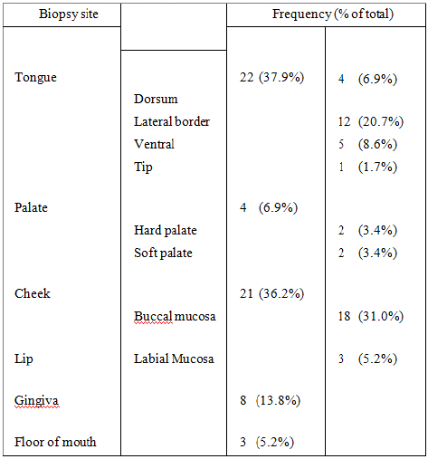
Figure 1: Table summarizing the region of each intra oral biopsy.
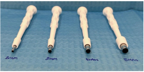
Figure 2: Various sizes of punch biopsies.
Mucosal biopsies accounted for 36.2% of all biopsies taken. The buccal mucosa accounted for the majority of the mucosal biopsies (85.7%), and 31.0% of total biopsies. The buccal mucosa was the next most frequent site to be assessed. The likely explanation is that the lateral border of the tongue & buccal mucosa are areas more commonly affected by trauma from mastication.
The size range of biopsy punches ranged from 2mm-6mm. The most commonly chosen size was 4mm for 27 biopsies (46.6%). A large size biopsy punch (6mm) was used on only one patient (1.7%). The table below breaks down the biopsies by the size of the punch, and shows the precise location.
Out of the total 58 biopsies, 57 (98.3%) of biopsies were diagnostic in the first instance. This did not meet the gold standard set of 100%. The biopsy which was diagnostically inadequate was a 2mm punch biopsy on the dorsum of the tongue and was cited on the histopathology report to have limited mucosa within the sample. An excisional biopsy of this area was then completed following on from the inconclusive result.
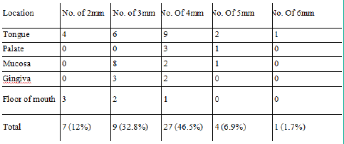
Figure 3: Table summarizing a further breakdown of chosen size of punch biopsy in each region of the mouth. All percentages have been rounded to the nearest 1 decimal place.
Two samples (3.4%), within the study had comments of artefact on the histopathological report. These have been recorded in the table below (Figure 4.). It should be noted that despite the inclusion of artefact in the histological sample, there was sufficient intact tissue to give an adequate diagnosis of the lesions.For most samples, histopathological results returned a benign diagnosis and patients were reassured by this fact. Within the study, three samples (5.2%) returned a confirmation of malignancy. These patients were then referred to and discussed at the head & neck oncology MDT meeting.
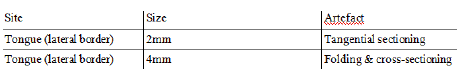
Figure 4: Table summarizing artifacts found on histological report.
Discussion
Our limited study shows that punch biopsies technique is a useful diagnostic tool for intra oral lesions. We are satisfied that on almost all occasions a correct & definitive histological diagnosis has been reached. Despite not meeting the gold standard of 100%, a diagnostic yield of 98.3% is much better than that reported by Gilvetti C et al [2], who showed sensitivity of 83% using a punch biopsy.
Moreover, in this study a punch biopsy is shown to be on par with other traditional biopsy methods, with a reported sensitivity of 53%. Gilvetti C et al [2] also suggested that a punch with a diameter of only 3mm is more reliable for achieving a diagnosis than a scalpel method. It should be noted that within our study, the single undiagnostic sample and half of the samples with reported artefact were carried out using a 2mm sample. This may suggest that operators should consider opting against using such a small punch biopsy, or for a complete excisional biopsy to ensure the sample is diagnostic and arrives to the histopathology lab with minimal artifact. These samples were also taken on the tongue, an area which can easily accommodate the use of a larger diameter punch.
Smaller diameter punches were typically used during gingival and floor of mouth biopsy, this may likely be due to the lesions being close to other anatomy in the area, and choice of a 2-3mm punch is less likely to cause damage to adjacent structures.
Within our study, a small number of biopsies returned a diagnosis of malignancy. This equated to 5.2% of the total sample size. This rate of malignant diagnosis from 2 week wait referrals of suspected oral cancer is in keeping with what has been reported previously in the literature by Rowlands CEP et al with an oral cancer conversion rate between 5-10%.
Identified limitations of our study include the single cycle of the audit and the relatively small sample size. Further cycles should be completed to ensure that the gold standard of diagnostic accuracy should be maintained and hopefully improved. As punch biopsies are so commonly used by junior members of the team within the department, we had no data to compare against the elliptical incisional biopsy technique. For a more adequate comparison between the two modalities, a prospective case control study could be completed alongside the next audit cycle. If this were to be completed, this should be accompanied by a teaching session for the junior members of the team on basic scalpel skills to try and mitigate any variables relating to the skill of the operator.
The traditional incisional method of biopsy requires an elliptical incision to be made via multiple cuts; the ideal length of the sample should be three times the length, and in an orientation which is along the grain of tissue fibres [5]. A punch biopsy is done using a disposable tool with a plastic handle & shaft, and a circular cutting blade with a range of sizes available from 2mm-10mm.
Optimal use of the punch biopsy tool is gained by initially applying perpendicular pressure to the lesion, and after this initial pressure using a twisting motion [2]. After using the punch, the deep margin of the sample is then released using scissors or a blade. When carrying out a biopsy, particularly for suspected malignancy, it is essential to include within the sample a small area of clinically normal looking tissue. It helps to identify the transition between the tissues [5]. In such instances, a larger punch size should be considered.
A systematic review and meta-analysis comparing the risk of artifact on a biopsy on an intra-oral lesion demonstrated that a punch biopsy is significantly less likely to produce tissue artefacts and as such should be the biopsy method of choice [3]. Scalpel biopsy, due to its multi-incision technique, can lead to the formation of multiple cuts in the specimen, which can also produce more localised bleeding [3]. Cuts & haemorrhage can lead to distortion artifact of the specimen and increase the risk of an inconclusive result, the design of a punch mitigates this risk.
A traditional wedge style incisional biopsy requires the specimen to be handled more due to more complex cuts and increased operative time when compared to the punch biopsy. The increased handling with tissue forceps can lead to compression of epithelial tissue into the underlying connective tissue, forming a pseudocyst like structure. The presence of pseudocysts can prevent accurate diagnosis of epithelial neoplasia [4]. The use of non-toothed forceps when handling the specimen has been advocated in several papers to reduce risk of artifact [2,3,5].
The diagnostic capability of an intra oral biopsy, time spent in the chair and pain from the procedure is all factors which greatly affect patients' experience. For junior members of staff, the procedure of a conventional intra-oral incisional biopsy, combined with the sometimes-awkward positioning of lesions, might cause difficulties. Punch biopsies offer a much more time effective and user-friendly technique for junior and experienced colleagues alike and also help reduce the incidence of sharp injuries to patient and clinician. Incisional biopsies tend to be larger in size than that of a punch biopsy – this means that the surgical site can take longer to heal which might leave patients in pain for much longer.
Despite punch biopsies generally being the quicker and safer method for intra-oral biopsy, there are some instances when a traditional scalpel method may be better indicated. Areas such as the maxillary tuberosity or lingual gingiva are difficult to reach using a biopsy punch, and in these instances a blade can be considered. Caution should also be used when using a punch biopsy on the floor of mouth due to the risk of damaging nearby structures, such as the Wharton duct, and subsequent risk of complication. However in the hands of an experienced and well-trained clinician the risks of damage to adjacent structure are equal to that of traditional methods, and as such a punch biopsy can still be suitable with or without the need for suture repair [2,3].
Conclusion
Punch biopsies are a quick and effective method of sampling intra oral lesions, which do not require specific surgical soft tissue skills. Punch biopsies are convenient and give sufficient tissue & depths and reduce risk of artefact on the biopsy, leaving a specimen which is suitable for histological diagnosis across a range of pathologies, leading to timely management for patients.
References
- Navone R, Burlo P, Pich A, Pentenero M, Broccoletti R, Marsico A, et al. The impact of liquid-based oral cytology on the diagnosis of oral squamous dysplasia and carcinoma. Cytopathology. 2007; 18: 356-60.
- Gilvetti C, Collyer J, Gulati A, Barrett AW. What is the optimal site and biopsy technique for the diagnosis of oral mucosal autoimmune blistering disease? J Oral Pathol Med. 2019; 48: 239-43.
- Rezende Oliveira S, Fernandes Araújo Almeida T, Aparecida da Silva T, Alves Mesquita R, Guimarães Abreu L. Comparison of tissue artifacts in punch and scalpel biopsies of oral and maxillofacial lesions: A systematic review and meta-analysis. J Stomatol Oral Maxillofac Surg. 2020; 121: 704-12.
- Margarone JE, Natiella JR, Vaughan CD. Artefacts in oral biopsy specimens. J Oral Maxillofac Surg. 1985; 43: 163-72.
- Oliver RJ, Sloan P, Pemberton MN. Oral Biopsies: methods and applications. Br Dent J. 2004; 196: 329-33.
- Singh D, Bastian TS, Singh A, Kudva S. Oral mucosal biopsy: comparison of surgical artefacts in incisional and punch oral mucosal biopsy. Medico-Legal Update. 2014; 14: 10-4.
- Rowlands CEP, James P, Lowe D, Rogers SN. Patient worry and concern associated with referral on the two-week suspected head and neck pathway. Br Dent J. 2022; 5: 1-5.