
Research Article
J Dent & Oral Disord. 2025; 11(1): 1190.
Use of Optical Coherence Tomography in the Detection of Microfractures in Teeth after Apicectomy: An In Vitro Comparative Study with Microct
Marcela Côrte Real Fernandes¹, Daniel Amancio Duarte², Márcio Fernando Paixão de Brito², Anderson Stevens Leonidas Gomes³, Antônio Celso Dantas Antonino², Andréa Cruz Câmara¹, José Antonio Poli de Figueiredo4* and Carlos Menezes Aguiar¹
¹Department of Prosthesis and Oral Surgery, Federal University of Pernambuco - UFP, Recife, Brazil
²Department of Nuclear Energy, Federal University of Pernambuco - UFP, Recife, Brazil
³Department of Physics, Federal University of Pernambuco - UFP, Recife, Brazil
4Department of Morphological Sciences, Federal University of Rio Grande do Sul - UFRGS, Porto Alegre, Brazil
*Corresponding author: José Antonio Poli de Figueiredo, Department of Morphological Sciences, Federal University of Rio Grande do Sul - UFRGS, Porto Alegre, Brazil Email: poli.figueiredo@outlook.com
Received: April 16, 2025 Accepted: April 26, 2025 Published: May 01, 2025
Abstract
Objectives: This study aimed to evaluate the ability of optical coherence tomography (OCT) to detect dentinal microcracks in teeth submitted to apicoectomy.
Methods: Forty-five extracted human permanent teeth were selected. After apicoectomy, specimens that had apical microcracks were identified using computed microtomography (μCT) as the gold standard. Then, the apical portions of the roots were scanned with spectral domain OCT (SD-OCT) and the resulting images were blindly evaluated by 3 independent examiners to detect microcracks. The diagnostic performance of the OCT device was calculated and statistical analysis was performed.
Results: Based on the OCT images, 17.8% (8) of the roots had dentine microcracks in the apical region. The images generated by the OCT were able to show lines of microcracks in the same location as the cross sections, however, it performed worse than the μCT because it demonstrated a limitation in the depth of the device, the images were not clear, and difficult to visualize for the observers.
Conclusion: The SD-OCT system presented low accuracy for detecting microfractures in teeth submitted to apicoectomy, compared to μCT. Clinical Significance: microCT still stands out as gold standard to detect microfractures, and caution should be taken when using OCT, as it does not display reliable images.
Keywords: Optical coherence tomography; microCT; Diagnosis; Tooth fractures; Apicoectomy
Introduction
The detection of incomplete fractures in the apical region of dental elements has been a diagnostic challenge for more than half a century due to the difficulty in discovering their location [1]. Some studies have observed the formation of root cracks in teeth submitted to apicoectomy and biomechanical preparation from channels [2-6].
Clinically, these microfractures have an obscure prognosis, as they can affect the healing process around the root, resulting in infiltration pathways, and leading to treatment failure [7]. Furthermore, they can spread, causing a vertical fracture, and can influence longterm survival. term of endodontically treated teeth [8-10]. Optical coherence tomography (OCT) is a non-invasive and non-destructive imaging technique, with relatively high resolution that allows obtaining and interpreting cross-sectional images of the research area [11-12]. It combines the principles of an ultrasound with the performance of a microscope image, using infrared light waves that reflect the internal microstructure within biological tissues [13]. It is worth mentioning the great potential of OCT in dentistry, having been reported to determine dentin thickness [14], in the detection of dentin microcracks in the apical portion [1], calculus on root surfaces [15], evaluation of caries [16], and dentin defects [17].
In clinical practice, some studies have evaluated the use of OCT to diagnose root fractures [18,19], however, the evidence is still insufficient. Given the above, the present study aimed to evaluate the accuracy of the SD-OCT system in detecting apical microfractures after apicoectomy. The null hypothesis tested was that OCT would perform similarly to microCT for detecting apical microfractures.
Materials and Methods
Sample Selection and Preparation
After approval by the Research Ethics Committee involving Human Beings of the Federal University of Pernambuco, Recife, Pernambuco, Brazil (CAAE: 49847221.3.0000.8808), 45 roots of human teeth, permanent, with a completed root formation process, carriers of single channels, confirmed radiographically. The specimens were obtained from the Human Teeth Bank of the Federal University of Pernambuco. Teeth were extracted for reasons unrelated to this study and stored in purified filtered water until use. The choice of roots was based on inspection and radiographic images in the mesiodistal and buccolingual direction, to exclude those with open apexes, endodontic treatment, dental calculus, root fractures, hypercementosis, internal/external resorption, and root caries.
To standardize the pre- and postoperative images and stabilize the roots, the specimens were included in a plaster block measuring 18x6 cm. Then, the blocks were numbered and randomly divided, by drawing lots, into three experimental groups according to the instrument to be used for the apicoectomy. In group 1, a truncatedconical diamond bur, n° 1064 (KG Sorensen, São Paulo, Brazil) was used; in 2, a surgical carbide drill n° 702 (Prima Dental by Angelus, Londrina/PR, Brazil) was used and in both groups, a high-speed pen was used. In group 3, a Bladesonic ultrasound tip (Helsé Ultrasonic) was used in the Ultrasound Jet Sonic BP – Gnatus with maximum power.
With the aid of an endodontic millimeter plastic ruler (Maquira, Maringá, Brazil), a distance of 3 mm below the apex of the root was marked with the aid of a permanent marker with a fine blue tip (Pilot pen, São Paulo, Brazil) and then the apicoectomy was performed along the long axis of the roots forming a 90° angle, with continuous water flow20.
Pre- and Post-Scanning of Specimens with Computed Microtomography
Using computed microtomography as the gold standard [1,21,22], the specimens were scanned before and after the apicoectomy procedure to identify microfractures. By image comparison method, they were identified in the apical portion of the roots by 3 independent observers.
The microtomograph used was XT H 225 ST INDUSTRIAL CT SCANNING SF, Nikon (Tokyo, Japan), with an integration time of 500 milliseconds per projection, voxel size of 10 micrometers, 100 kV, and 100 μA with a 1 mm thick aluminum filter. Each root was mounted on a metallic table, which is an integral part of the tomograph, and scanned whose acquisition of specimen projections was performed at different angles along a 360° rotation with a voxel size of 10 μm and a time of acquisition of 25 minutes, generating 3,016 projections of each scan.
To reduce ring artifacts and minimize the effect of beam hardening, an air calibration of the detector was performed before the scans. Images of each specimen were reconstructed using CT Pro 3D software v.XT3.13 (Nikon Metrology, NY, Tring, UK). Then, VG Studio Max 2.2 software (Volume Graphics, Heidelberg, Germany) was used to smooth images by applying a Gauss filter and providing cross-sections of the internal structure of the roots.
Scanning with Optical Coherence Tomography (OCT)
After apicoectomy, the specimens were scanned using the commercial OCT system (Callisto, Thorlabs, Inc., New Jersey, USA), operating in a spectral domain (SD-OCT) that employs a super luminescent diode as a light source with 930 nm of central wavelength, 100 nm of spectral bandwidth and maximum output power of 5mW. The axial resolution of the 6.0 μm system is determined by the light source bandwidth, while the geometric characteristics of the focused beam are related to the lateral resolution.
The system consists of three main parts: a handpiece, a base unit, and a computer. The base unit contains the light source, which employs the super luminescent diode and the light is directed through an optical fiber, which is located inside the handpiece. For imaging, specimens were placed on the worktable with the apex facing upwards and the scanning light beam was oriented parallel to the long axis of the teeth above the root apex. With the use of an infrared detector (FIND-R-SCOPE, Model 84499C-5), the light beam was positioned at the most central point of the apex, and, from this point, a scan was performed in the mesiodistal and buccolingual direction of all specimens. Two images are captured per second, one in the horizontal direction and the other in the vertical direction of the specimens with a resolution of 6 μm in depth and 10 μm in width per pixel. Regarding depth, the device allows a maximum penetration of approximately 1.7 mm into the image.
The entire system was controlled by software compatible with LabVIEW that collects and processes the data, generating the tomographic image.
Evaluation of Microcracks
Three independent examiners blindly evaluated two images of each specimen, one in the horizontal direction and the other in the vertical direction, produced by OCT obtained from the apical portion of the roots. The examiners were specialists in endodontics previously trained with OCT images generated from teeth not included in the study sample. Each examiner scored the presence or absence of microfractures on the teeth according to a 5-point scale: 1, definitely absent; 2, probably absent; 3, uncertain; 4, probably present; and 5, definitely present. The images were reassessed after an interval of 15 days.
Statistical Analysis
The results obtained from the microcrack evaluation of the SDOCT device were compared with the gold standard (microCT). Sensitivity, specificity, positive predictive value, negative predictive value, and accuracy were determined for each device and analyzed using Fisher's exact test. Furthermore, the ROC curve was calculated and the areas under the ROC curves were compared using the DeLong test. To assess inter-examiner and intra-examiner agreement, the weighted Kappa test was used. Data were analyzed using SPSS v.23 statistical software (SPSS Inc. Chicago, IL) and MedCalc for Windows v.14.8.1 (MedCalc Software BVBA, Ostend, Belgium). The significance level was 5%.
Results
Based on the OCT images, it was found that 17.8% (8) of the roots had dentin microcracks after apicoectomy. The cross-sectional images produced by microCT and SD-OCT scanning are shown in Figures 1 and 2.
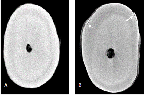
    Figure 1: Images of specimens performed with computed
microtomography. (A) a specimen without detection of fractures before
apicoectomy; (B) a specimen with fracture detection (arrow).
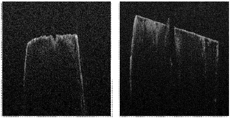
    Figure 2: Images with the SD-OCT device. (A) a specimen without
detection of fractures before apicoectomy; (B) a specimen with fracture
detection (arrow).
Table 1 shows the values of sensitivity, specificity, positive prediction, negative predictive value, precision, and area under the ROC curve in which the OCT device presented an inferior diagnostic performance for all evaluated parameters.
![]()
    Statistical    parameter
    OCT


    Sensitivity
    33.33%


    Specificity
    71.30%


    PPV
    22.50%


    NPV
    81.05%


    Accuracy
    63.70%


    AUC
    52.30%


    PPV: Positive Predictive Value; NPV: Negative    Predictive Value; AUC: Area Under ROC Curve
Table 1: Diagnostic performance of OCT.
Discussion
In the present study, optical coherence tomography (OCT) was chosen because it is a non-invasive technique and provides crosssectional images of a tooth structure [23]. It is a promising imaging modality for visualizing dental cavities [24], dental fractures [5,25,26], interfacial gaps in restorations [17], and root canals [13], among other situations in dentistry. One of the great advantages is the use of light instead of radiation, presenting itself as a safer alternative due to the absence of x-rays, minimizing risks for the operator [22,27,28].
The present study demonstrated that the SD-OCT device detected microfractures in teeth submitted to apicoectomy, however, when compared to computed microtomography, its performance was inferior. Particularly, a sensitivity of 33.33% may be due to the darkness of the image provided, or lack of familiarity with OCT from the observers. The use of resources to improve the images may help this in future analyses.
Computed microtomography provides a three-dimensional, non-destructive image of the specimens, and successive scans can be performed after different treatments [29]. In OCT, crosssectional images are generated by multiple axial axes. The equipment technique uses infrared light waves that are reflected in the internal microstructure in a way that, in principle, is analogous to an ultrasonic pulse-echo [11].
The difficulty reported by the observers, when viewing the image, is correlated with the maximum penetration of the device, which in the study allowed a maximum penetration of 1.7 mm. The apical resection occurred within 3 mm from the apical portion, so when comparing the pre- and post-apicoectomy image, the comparison region was not as evident as in the microCT.
In one study, the OCT device was able to distinguish structural cracks, lines of microcracks, and lines of division in cracks in teeth [30]. In the present study, when evaluating the apical region, the dental structures were not distinguished and all the microcracks appeared as dark cracks separating the dentin.
With regard to the apicoectomy technique, they recommend that the method be highly refined to favor the controlled removal of the root apex and produce smooth and clean surfaces, thus avoiding excavations or the sulcus effect [31]. In the present study, the difficulty in detecting microcracks also correlates with the type of instrument used in the apicoectomy, where the lack of surface smoothness may have contributed to a false diagnosis in the image produced by the OCT. Grooves and excavations formed on the surface appeared as a crack in the dentin region and due to the depth limit of the device, the defects were interpreted as microcracks.
Within the limitations of this laboratory study, as a diagnostic method for microfractures in teeth submitted to apicoectomy, the OCT device presented low accuracy compared to μCT. Even though OCT has the advantage of in vivo measurements, a deeper penetration depth, or improvement in the image processing, may be required for a better use of this device compared to what was reported in this study.
Funding
This research did not receive any specific grant from funding agencies in the public, commercial, or not-for-profit sectors. This study was partially supported by grants from FACEPE, CAPES and CNPq, Brazilian agencies.
Acknowledgments
The authors deny any conflicts of interest related to this study. This study was partially supported by grants from FACEPE, CAPES and CNPq, Brazilian agencies.
References
- de Oliveira BP, Câmara AC, Duarte DA, Gomes ASL, Heck RJ, Antonino ACD, et al. Detection of Apical Root Cracks Using Spectral Domain and Sweptsource Optical Coherence Tomography. J Endod. 2017; 43: 1148–1151.
- Aydemir S, Cimilli H, Mumcu G, Chandler N, Kartal N. Crack formation on resected root surfaces subjected to conventional, ultrasonic, and laser rootend cavity preparation. Photomed Laser Surg. 2014; 32: 351–355.
- Beling KL, Marshall JG, Morgan LA, Baumgartner JC. Evaluation for cracks associated with ultrasonic root-end preparation of Gutta-Percha filled canals. J Endod. 1997; 23: 323–326.
- Layton CA, Marshall JG, Morgan LA, Baumgartner JC. Evaluation for cracks associated with ultrasonic root-end preparation. J Endod. 1996; 22: 157-160.
- Russell AA, Chandler NP, Friedlander LT. Crack formation following root-end preparation in roots with the butterfly effect. Eur Endod J. 2018; 3: 107–112.
- Saunders WP, Saunders EM, Gutmann JL. Ultrasonic root-end preparation Part 2, Microleakage of EBA root-end fillings. Int Endod J. 1994; 27: 325-329.
- Ayranci F, Ayranci LB, Özdogan A, Ozkan S, Peker MO. Aras MH. Resistance to vertical root fracture of apicoected teeth using different devices during two root canal irrigation procedures. Lasers Med Sci. 2018; 33: 1685–1691.
- Von Arx T, Steiner RG, Tay FR. Apical surgery: Endoscopic findings at the resection level of 168 consecutively treated roots. Int Endod J. 2011; 44: 290–302.
- Yan W, Montoya C, Øilo M, Ossa A, Paranjpe A, Zhang H, et al. Contribution of Root Canal Treatment to the Fracture Resistance of Dentin. J Endod. 2019; 45: 189–193.
- Çapar ID, Gök T, Uysal B, Keles A. Comparison of microcomputed tomography, cone beam tomography, stereomicroscopy, and scanning electron microscopy techniques for detection of microcracks on root dentin and effect of different apical sizes on microcrack formation. Microsc Res Tech. 2019; 82: 1748–1755.
- Huan D, Swanson EA, Lin CP, Schuman JS, Stinson WG, Chang W, et al. Optical Coherence Tomography. Sci Rep. 1991; 254: 1178–1181.
- Podoleanu AG. Optical coherence tomography. J Microsc. 2012; 247: 209– 219.
- Shemesh H, Van Soest G, Wu MK, Van Der Sluis LWM, Wesselink PR. The Ability of Optical Coherence Tomography to Characterize the Root Canal Walls. J Endod. 2007; 33: 1369–1373.
- Majkut P, Sadr A, Shimada Y, Sumi Y Tagami J. Validation of Optical Coherence Tomography against Micro-computed Tomography for Evaluation of Remaining Coronal Dentin Thickness. J Endod. 2015; 41: 1349–1352.
- Krause F, Schmalz G, Park KJ, Schmidt J, Zielolz D, Schneider H, Haak R. Evaluation of calculus imaging on root surfaces by spectral-domain optical coherence tomography. Photodiagnosis Photodyn Ther. 2019; 25: 275–279.
- Shimada Y, Yoshiyama M, Tagami J, Sumi Y. Evaluation of dental caries, tooth crack, and age-related changes in tooth structure using optical coherence tomography. Jpn Dent Sci. 2020; 56: 109-118.
- Bakhsh TA, Sadr A, Shimada Y, Tagami J, Sumi Y. Non-invasive quantification of resin-dentin interfacial gaps using optical coherence tomography: Validation against confocal microscopy. Dent Mater. 2011; 27: 915–925.
- Shemesh H, Van Soest G, Wu MK, Wesselink PR. Diagnosis of Vertical Root Fractures with Optical Coherence Tomography. J Endod. 2008; 34: 739–742.
- Yoshioka T, Sakaue H, Ishimura H, Ebihara A. Suda H. Sumi Y. Detection of root surface fractures with swept-source optical coherence tomography (SSOCT). Photomed Laser Surg. 2013; 31: 23–27.
- Kim S. Principles of endodontic microsurgery. Dent Clin North Am. 1997; 41: 481–497.
- De-Deus G, Belladonna FG, Marins JR, Silva EJNL, Neves AA, Souza EM, et al. On the causality between dentinal defects and root canal preparation: A Micro-CT assessment. Braz Dent J. 2016; 27: 664–669.
- Rashed B, Lino Y, Ebihara A, Okiji T. Evaluation of Crack Formation and Propagation with Ultrasonic Root-End Preparation and Obturation Using a Digital Microscope and Optical Coherence Tomography. 2019; 1-6.
- Hsieh YS, Ho YC, Lee SY, Chuang CC, Tsai J, Lin KF, Sun CW. Dental optical coherence tomography. 2013; 13: 8928–8949.
- Trebing CT, Schwindling FS, Leisner L, Trebing J, Lux CJ, Rammelsberg P, Sen S. Diagnostic accuracy of 870-nm spectral-domain OCT with enhanced depth imaging for the detection of caries beneath ceramics. J Dent. 2020; 102: 1-7.
- Lee SH, Lee JJ, Chung HJ, Park JT, Kim HJ. Dental optical coherence tomography: new potential diagnostic system for cracked-tooth syndrome. Surg Radiol Anat. 2016; 38: 49–54.
- Braz AKS, Kyotoku BBC, Braz R, Gomes ASL. Evaluation of crack propagation in dental composites by optical coherence tomography. Dent Mater. 2009; 25: 74–79.
- de Melo LSA, de Araujo RE, Freitas AZ, Zezell D, Vieira JrND, Girkin J, et al. Evaluation of enamel dental restoration interface by optical coherence tomography. J Biomed Opt. 2005; 10.
- Shimada Y, Sadr A, Sumi Y, Tagami J. Application of Optical Coherence Tomography (OCT) for Diagnosis of Caries, Cracks, and Defects of Restorations. Curr Oral Health Rep. 2015; 2: 73–80.
- Campello AF, Marceliano-Alves MF, Provenzano JC, Loyola SC, Siqueira JrJF, Machado AG, et al. Accuracy of Microcomputed Tomography in Detecting Dentinal Cracks: A Correlative Study with Scanning Electron and Operative Microscopy. Scanning. 2021; 1-7.
- Kim JM, Kang SR, Yi WJ. Automatic detection of tooth cracks in optical coherence tomography images. J Periodontal Implant Sci. 2017; 47: 41–50.
- Duarte MAH, Domingues R, Matsumoto MA, Padovan LEM, Kuga MCK. Evaluation of apical surface roughness after root resection: a scanning electron microscopic study. Oral Surg Oral Med Oral Pathol Oral Radiol Endod. 2007; 74-76.
Citation:Marcela Côrte Real Fernandes, Daniel Amancio Duarte, Márcio Fernando Paixão de Brito, Anderson Stevens Leonidas Gomes, Antônio Celso Dantas Antonino, et al. Use of Optical Coherence Tomography in the Detection of Microfractures in Teeth after Apicectomy: An In Vitro Comparative Study with Microct. J Dent & Oral Disord. 2025; 11(1): 1190.