Clinical Image
Austin J Clin Ophthalmol. 2022; 9(3): 1133.
Cerulean Cataract
Belidi HE*, Saoiabi Y, Boumehdi I and Cherkaoui O
Department of Ophthalmology, Hopitaldes Spécialités, Rabat, Morocco
*Corresponding author: Belidi HE, Department of Ophthalmology, Hopitaldes Spécialités, Rabat, Morocco
Received: October 25, 2022; Accepted: November 09, 2022; Published: November 16, 2022
Clinical Image
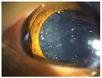
A 14-year-old girl presented with a two years history of gradual decrease of vision in both eyes. The best corrected visual acuity was 0, 3 LogMARin both eyes. The examination of the anterior segment on slit-lamp of both eyes releveled multiple tiny bluish-white opacities distributed in the lensnucleus and cortexin the form of concentric circles corresponding to a congenital cerulean cataract. No other abnormality was observed in the slit lamp in both eyes. Phacoemulsification surgery was planned for each eye with a good evolution.

Figure 1: Diffuse light slit lamp photograph of the left eye showing multiplebluish-white opacities spread throughout the cortex of lens.
Cerulean cataract, also known as blue dot cataract, is a rare phenotypic variant of congenital cataract, first described by Vogt [1]. Cerulean cataracts are inherited as an autosomal dominant trait [2]. It is a developmental cataract characterized by bluish-white opacifications scattered in the nucleus and cortex of the lens [3]. Patients are usually asymptomatic until the age of 18–24 month.
Disclosure of Interest
The authors declare that they have no competing interest.
References
- Francis PJ, Berry V, Bhattacharya SS, Moore AT. The genetics of childhood cataract. J Med Genet. 2000; 37: 481–8.
- Litt M, Carrero-Valenzuela R, LaMorticella DM, et al. Autosomal dominant cerulean cataract is associated with a chain termination mutation in the human beta-crystallin gene CRYBB2. Hum Mol Genet. 1997; 6: 665–8.
- Ram J, Singh A. Cerulean cataract. QJM. 2019; 37.