Clinical Image
Austin J Clin Ophthalmol. 2017; 4(1): 1079.
Cilioretinal Artery Sparing Central Retinal Artery Occlusion
S Pirasath¹, N Suganthan², M Malaravan³
¹Registrar, 2Consultant Physician and Senior Lecturer in Clinical Medicine, Professorial Medical Unit, 3Consultant Ophthalmologist, Teaching Hospital, Jaffna, Sri Lanka
*Corresponding author: Selladurai Pirasath, Registrar in Clinical Medicine, Professorial Medical Unit, Teaching Hospial, Jaffna
Received: June 05, 2017; Accepted: June 13, 2017; Published: June 20, 2017
Keywords
Retinal artery occlusion; Cilioretinal artery
Clinical Image
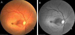
A 55 year-old male presented with a history of sudden onset of vision loss on his right eye, lasting for 24 hours duration on a background of Hypertension. Further, he had right side ischemic stroke a year ago. On clinical examination, best-corrected visual acuity (BCVA) was hand movement and 6/9 in right and left eye respectively. Fundus examination revealed right eye central retinal artery occlusion with sparing cilioretinal artery (Figure 1A & 1B). His cardiovascular examination revealed irregularly irregular pulse with a heart rate of 140 beats per minutes and blood pressure of 110/70mmHg. His Barthel Index of Activities of Daily Living was 14. Subsequently ECG confirmed the diagnosis of atrial fibrillation with the CHA2DS2-VASc Score of 4. His basic biochemical and hematological investigations including ESR, C-reactive protein (CRP), and platelet levels were normal. He was started on warfarin with a target INR of between 2 and 3. On assessment at one month, he showed slight improvement on his best corrected vision of 6/60 in right eye.

    Figure 1:  1A & 1B: The fundal photograph of right eye showed oedmatous and
opaque except the cilioretinal artery territory supply indicating central retinal
artery occlusion with cilioretinal artery sparing.
