
Special Article - Ophthalmology: Clinical Cases and Images
Austin J Clin Ophthalmol. 2016; 3(2): 1069.
Imitator of the Conjunctivitis: Bilateral Posterior Scleritis in a Child Patient
Resat Duman, Rahmi Duman*, Sibel İnan and Mustafa Doğan
Department of Ophthalmology, School of Medicine, Afyon Kocatepe University, Turkey
*Corresponding author: Duman R, Department of Ophthalmology, School of Medicine, Afyon Kocatepe University, Ali Çetinkaya Kampusü, Izmir Karayolu 7. Km, Afyonkarahisar, Turkey
Received: June 07, 2016; Accepted: July 19, 2016; Published: July 25, 2016
Abstract
Conjunctivitis, keratitis, uveitis, episcleritis and scleritis are the most common causes of red eye. Painful red eye is the most common presentation of Posterior Scleritis (PS). PS rarely occurs in childhood. Herein, we report a 13-year-old male who presented with visual loss, pain and redness. Following B-scan ultrasonography and optical coherence tomography examination of the eyes, a diagnosis of posterior scleritis was made, and the patient was successfully treated with oral steroid therapy.
Keywords: Posterior scleritis; Conjunctivitis; Ultrasonography
Introduction
Conjunctivitis, keratitis, uveitis, episcleritis and scleritis are the most common causes of red eye. Painful red eye is the most common presentation of Posterior Scleritis (PS) [1]. Symptoms of PS include particular pain, blurred vision and photophobia. Posterior scleritis is a rare condition and generally associated with different systemic diseases such as rheumatoid arthritis and polyarthritis nodosa [2]. The rate of detectable sign of PS on the first examination is 83% [3]. The mean age at onset is 49 years [4]. PS occurrences in children are very rare and the ophthalmic literature consists of predominantly single case reports. 5Wepresent a child case with posterior scleritis.
Case Presentation
A 13-year-old boy presented with a 1monthhistory of bilateral visual loss, pain and redness. He had been entreated with topical antibiotic and steroid with a diagnosis of conjunctivitis and uveitis previously. He had no history of arthralgia, fever, cough, malaise or weight loss and any systemic disease. Patient underwent a complete ophthalmological examination, including fluoresces in angiography, B-scan ultrasonography and optical coherence tomography. In addition, physical and laboratory examination including serologic analysis for systemic diseases were performed.
Proptosis was not observed and ocular motility was normal. Intraocular pressures were normal bilaterally. No evidence of relative afferent pupillary defect was noted. Chemosis was present in both eyes. The visual acuities of the right and left eyes were 2/10 and 5/10, respectively initially. The corneas were clear with showed 2+ cells in the anterior chamber. Fund us photography of the right eye showed a round-shaped choroidal mass of about three disc diameters in size with choroidal folds located in the inferotemporal quadrant. Bilateral optic discs were hyperemic and increased tortuosities of the vessels were present in the right eye (Figure 1). B-scan ultrasonography demonstrated choroidal and scleral thickening and a classic T sign with increased acoustic density of the choroid and sclera bilaterally (Figure 2).
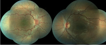
    Figure 1:  Fundus photography of the right eye showed a round-shaped
choroidal mass of about three disc diameters in sizewith choroidal foldslocated
in the inferotemporal quadrant.
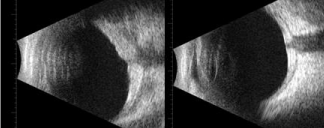
    Figure 2:  B-scan ultrasonography demonstrated choroidal and scleral
thickening and a classic T sign with increased acoustic density of the choroid
and sclera in our patient bilaterally.
Optical coherence tomography showed exudative retinal detachment around the optic disc bilaterally and inferotemporal quadrant in the right eye (Figure 3).
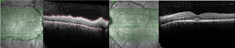
Figure 3: Optical coherence tomography showed exudative retinal detachment around the optic disc bilaterally and inferotemporal quadrant in the right eye.
Neuroimaging with Magnetic Resonance Imaging (MRI) of the head and orbits revealed normal findings. When the Intravenous Fundus Fluoresce in Angiography (FFA) assessed; increased hyper fluorescense in the late periods was remarkable bilaterally optical discs. It was determined that there are no leaks from the vessels early and late periods (Figure 4).
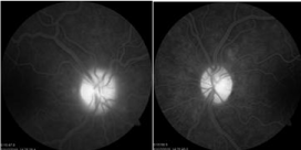
    Figure 4:  It was determined that there are no leaks from the vessels early
and late periods and increased hyperfluorescense in the late periods was
remarkable bilaterally optical discs.
The patient was referred to a pediatrician for further evaluation. The labaratory examination, including a complete blood count, liver function, renal function, syphilis, toxoplasma serology, antinuclear antibodies, anti DNA and rheumatoid factor gave results within normal limits. There was an increase in C Reactive Protein (CRP) of 2, 28 mg/dL (0-0.8mg/dL). However, he was not diagnosed with any autoimmune disease. With these findings, a diagnosis of posterior scleritis was made.
Our patient was followed for 5 days with topical steroid treatment and non-corticosteroid anti inflammatory medication. On the basis of these clinical findings and after being ruled out infectious causes, a diagnosis of PS was made and the patients were treated with oral steroids (1 mg/kg). Patients during follow-up; 2 month was completely cutoff by reducing the dose of oral steroids.
Three weeks after initiation of therapy, chemosis had resolved bilaterally. But mild choroidal effusions remained in the right eye. Two months after therapy, BCVA improved to 10/10 bilaterally. With complete resolution of chemosis and choroidal lesions in both eyes.
Discussion
Posterior scleritis is a potentially sight-threatening rare condition that estimated to account for only 2% to 12% of all cases [5]. PS in adults is twice as common in women as in men; however males are affected more than females in children [5]. Posteriorscleritis in children is even rarer than in the adult population. The age distribution of patients with PS is 7 months to 19 years (median 12 years).
A typical clinical presentation is characterized by a range of clinical findings, like as anterior segment inflammation (64.8%), discs welling (79.6%), choroidal folds, retinal striaorserous retinal detachment (64.8). 4Bilateral involvement at presentation is frequent (53.8%) [5].
The diagnosis of PS is difficult with a quiet anterior segment. Fluoresce in angiography, orbital MRI, B-scan USG and OCT may be helpful. T-sign (the presence of fluid in the sub-Tenon’s space) is the most useful confirmatory ultrasound finding sign in patients with posterior scleritis.
Idiopathic central serous chorioretinopathy, traumatic retinal pigment epitheliopathy, and neoplastic infiltration of the choroid must be considered in the differential diagnoses. Exudative retinal detachments with choroidal masses in children should suggest infiltration with leukemia or lymphoma cells [6-7]. Complete blood count and orbital MRI (visualize hyper intensity around the globe, indicating an inflammatory process in PS) are important in differential diagnosis. Patients should assess by the pediatrician. Idiopathic Central Serous Chorioretinopathy (ISSR) in childrenery rare entity [8]. Differential diagnoses distinct from (ISSR) is made by the angiographic appearance of numerous pinpoint leaks with late staining of the optic nerve head and the thickening of the sclera and choroid on ultrasonography. These findings were not consistent with the diagnosis of idiopathic central serous chorioretinopathy.
The patient in our study appeared to have posterior scleritis with the clinical features included ocular pain with decreased visual acuity, and a thickened choroid and sclera with high acoustic density on ultrasonography.
Systemic collagen vascular diseases like rheumatoid arthritis, systemic lupus erythematosus and wegener’s granulomatosis are important associations in adults however it has not been associated with any systemic autoimmune disease in children until now. 5 In our patient any systemic disease were not detected too.
Posterior scleritis in children was generally resolved with oral systemic corticosteroids and non-steroidal anti-inflammatory drugs [9-10]. Our patient received two months of oral corticosteroids with good response. The risk of relapse in PS is high with an incidence rate of 15.81% per person/year [4].
The complaint of eye pain, the thickening of the sclera and choroid on ultrasonography, FFA and OCT may provide clues to the diagnosis of this rare disease.
References
- Watson PG, Hayreh SS, Awdry PN. Episcleritis, Scleritis I. Br J Ophthalmol. 1968; 52: 278-279.
- Liu AT, Fiona OL, Carmen KC. A case of giant nodular posterior scleritis mimicking choroidal malignancy. Indian journal of ophthalmology. 2015; 63: 919-921.
- Mc Cluskey PJ, Watson PG, Lightman S, Haybittle J, Restori M, Branley M. Posterior scleritis: Clinical features, systemic associations, and outcome in a large series of patients. Ophthalmology. 1999; 106: 2380-2386.
- Lavric A, Gonzalez-Lopez JJ, Majumder PD, Agrawal R. Posterior the thickening of the sclera and choroid on ultrasonography Scleritis: Analysis of Epidemiology, Clinical Factors, and Risk of Recurrence in a Cohort of 114 Patients. Ocular immunology and inflammation. 2015; 24: 1-10.
- Cheung, CMG, Chee SP. Posterior scleritis in children: clinical features and treatment. Ophthalmology. 2012; 119: 59-65.
- Burns CA, Blodi FC, Williamson BK. Acute lymphocytic leukemia and central serous retinopathy. Trans. Am. Acad. Ophthalmol. Otolaryngol. 1965; 69: 307-309.
- Zimmerman, LE, Thoreson HT. Sudden loss of vision in acute leukemia. A clinicopathologic report of two unusual cases. Surv. Ophthalmol. 1964; 9: 467-473.
- Fine SL, Owens SL. Central serous retinopathy in a 7-year-old girl. Am. J. Ophthalmol.1980; 90: 871-873.
- Hage R, Jean‑Charles A, Guyomarch J, Rahimian O, Donnio A, Merle H. Nodular posterior scleritis mimicking choroidal metastasis: A report of two cases. ClinOphthalmol. 2011; 5: 877‑880.
- Sridharan S, Juneja R, Hussain A, Biswas J. Giant nodular posterior scleritis mimicking choroidal tumor. Retin Cases Brief Rep. 2007; 1: 65‑67.