Clinical Image
Austin J Clin Ophthalmol. 2015; 2(5): 1059.
Cilioretinal Artery Sparing Central Retinal Artery Occlusion
Mathew DJ*, Sarma SK and Basaiawmoit JV
Department of Ophthalmology, Bansara Eye Care Centre, India
*Corresponding author: Mathew DJ, Department of Ophthalmology, Bansara Eye Care Centre, Laitumkhrah, Shillong, India
Received: November 06, 2015; Accepted: December 02, 2015; Published: December 11, 2015
Clinical Image
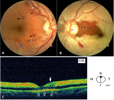
A gentleman aged 53 years who was a hypertensive on irregular medication presented with decreased vision in the left eye for the last 24 hours. His best corrected visual acuity was 20/30 and counting fingers close to the face in the right and left eye respectively. His blood pressure was 230/110 mm Hg at presentation. Glycosylated haemoglobin (HbA1c) was 8.8%. Right eye fundus showed haemorrhages, hard exudates, arteriolar attenuation and arteriovenous crossing changes suggestive of hypertensive retinopathy (Figure 1A). Left eye fundus showed white opacification of the retina except for a small island between the disc and the fovea supplied by the cilioretinal artery (Figure 1B). As it was already 24 hours after the onset of symptoms no intervention was undertaken for the CRAO. OCT line scan directed from inferior to superior showed typical inner retinal layer hyperreflectivity with thickening and outer retinal layer hypo-reflectivity sparing the fovea and a small area of superior perifoveal retina supplied by the cilioretinal artery (Figure 1C). After giant cell arteritis was ruled out with a normal ESR, he was referred to a cardiologist for further evaluation and management. He did not have any sources of embolism on evaluation. Ten days later, vision in the left eye was 20/80 eccentrically which stabilized thereafter.

    Figure 1:  A) Right eye fundus showing haemorrhages (black solid arrows),
hard exudates (blue arrows), arteriolar attenuation (black hollow arrows) and
arteriovenous crossing changes (white arrow) suggestive of hypertensive
retinopathy.
B) Left eye fundus showing whitening of the retina except for a small island
between the disc and the fovea supplied by the cilioretinal artery (black hollow
arrows)
C) OCT line scan directed from inferior to superior showing inner retinal
layer hyper-reflectivity with thickening and outer retinal layer hypo-reflectivity
sparing the fovea and a small area of superior perifoveal retina (white solid
arrow) supplied by the cilioretinal artery. The normal outer retinal layer hyperreflectivity
is seen clearly at the fovea and the spared region (white hollow
arrows).