Case Report
Austin J Clin Ophthalmol. 2015;2(3): 1051.
Pars Plana Vitrectomy, Lensectomy, Stalk Transection with Capsulectomy for the Primary Treatment of Persistent Fetal Vasculature Cataract
Nelson JW¹, Bach A¹, Villegas VM², Gold AS²,Wildner AC², Thompson J², Latiff A² and Murray TG²*
1Larkin Community Hospital – 7031 SW 62nd Avenue South Miami, USA
2Murray Ocular Oncology & Retina, 6705 Red Road, Suite 412, Miami, USA
*Corresponding author: Timothy G. Murray, Murray Ocular Oncology & Retina, 6705 Red Road, Suite 412,Miami, FL 33143, USA
Received: March 18, 2015; Accepted: June 17, 2015; Published: June 23, 2015
Abstract
Purpose: The purpose of this report is to present an alternative surgical approach to persistent fetal vasculature.
Case: A 9 month old male presented with leukocoria in the right eye and was found to have lenticular opacity with posterior dysplastic mass with complex exudative tractional retinal detachment and fibrovascular stalk. Surgical approach consisted of fibrovascular membrane transection anterior to termination of the stalk. This was performed without endodiathermy despite vascularization. The posterior capsule was opened and stalk was removed. Lensectomy was performed to remove lens nucleus and cortex. The patient was left aphakic and the both anterior and posterior capsules were opened widely. One month follow up of the patient showed a clear visual axis and refractive error of +5.00 spheres OD
Discussion: Surgical morbidity remains a major hurdle in the management of persistent fetal vasculature. The use of endodiathermy has gained popularity, but has the disadvantage of introducing intraocular energy. There has been a lack of literature regarding the management of persistent fetal vasculature with the presence of posterior involvement, and this case presents an alternative approach without the need for endodiathermy despite vascularization.
Keywords: Persistent fetal vasculature; Persistent hyperplastic primary vitreous; Pediatric ophthalmology
Introduction
Persistent Fetal Vasculature (PFV), a pathology first described by Reese as persistent hyperplastic primary vitreous [1], results from failure to in volute the primary vitreous and hyaloids vasculature during the 2nd trimester. The most classic presentation results in a persistent vascular stalk extending from the optic disc to the posterior lenticular capsule. However, significant variability in the spectrum of this disease has been reported [2-5]. PFV is most often unilateral and is associated with decreased axial length, hyperopia, cataract, and glaucoma [6-9].
Surgical management of patient with PFV remains a challenge. Significant complications including posterior synechiae, retinal detachment, phthisis, posterior capsular opacity, inflammatory pupillary membrane, glaucoma, intraocular lens displacement, and vitreous hemorrhage have been reported [8,10-12]. Despite these complications, post-operative improvement in visual acuity has been reported as high as 83% [8]. Various surgical techniques have been documented [3,8,10,11,13-18], however the literature regarding posterior segment surgical approach remains scarce. We report a patient that underwent pars plana vitrectomy, lensectomy, and posterior capsulectomywithout the use of endodiathermy as the primary treatment for cataract associated to PFV.
Case Report
A nine-month-old boy born at term via uncomplicated cesarean section was evaluated due to leukocoria in his right eye (OD). The mother had routine prenatal care with an uneventful pregnancy, and no other pertinent medical or surgical history.
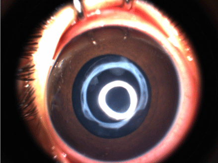
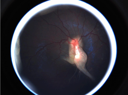
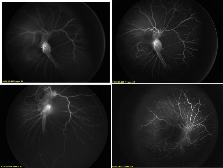
Ophthalmologic examination was remarkable for decreased visual acuity as evidenced by poor fixation and exotropia OD. There was no relative afferent pupillary defect and intraocular pressure (IOP) was 9 mmHg in both eyes (OU). Anterior segment examination was unremarkable OU, with the exception of a sector a cataract OD. (Figure 1). Indirect ophthalmoscopy was remarkable for a preretinal posterior dysplastic mass with complex exudative tractional retinal detachment and a fibrovascular stalk OD (Figure 2). There were no macular holes, tears, or breaks OU. Vascular ectatic alterations, as seen in fluorescein angiography (Figures 3a & 3b), were present OU. Due to the presence of a visually significant cataract, the patient was scheduled for pars plana vitrectomy, and lensectomy with capsulectomy as described below:

    Figure 1:  Sectoral Lenticular Cataract, right eye, in patient with persistent
fetal vasculature.

    Figure 2:  Fundus photo, right eye, showing preretinal posterior dysplastic
mass with complex exudative tractional retinal detachment and fibrovascular
stalk.

    Figure 3:  Vascular ectatic alterations with vascularized stalk, right eye, seen
on fluorescein angiography.
The right eye was prepped and draped and a wire lid speculum was inserted. The 25 gauge valved trocars were introduced 1.5 mm posterior to the limbus and posterior to the lens. The infusion line was then attached and IOP was stabilized. The primary 25 gauge vitrector and endoilluminator were introduced and, with contact 130 degree wide field viewing, a primary vitrectomy was performed. There were marked tractional alterations encircling the membranous stalk. There was posterior retinal dysplasia. The termination of the stalk was identified and, anterior to this, a transection of the fibrovascular membrane was performed. There was direct lenticular insertion of the stalk with lenticular alterations. The posterior capsule was then opened, and the stalk was removed in its entirety. Lensectomy techniques were utilized to remove all of the lens nucleus and cortex. Due to the patient’s age and a crowded anterior segment, it was determined appropriate to leave the patient aphakic. The capsule was then opened both anterior and posterior widely to deal with the potential postsurgical capsular phimosis. After the procedure was completed, the retina was noted to be free of open holes, tears, or breaks. The patient received subconjunctival injection with dexamethasone and gentamicin 180 degrees away from the valved trocars. Topical prednisolone acetate and gentamicin were also applied followed by an occlusive patch and Fox shield.
Follow up evaluation at one month showed a clear visual axis, a refractive error of +5.00 sphere OD, and restoration of the posterior foveal anatomy and vascular sclerotic alterations. There was no active retinal hole, tear, break, epiretinal membrane, or proliferative vitreoretinopathy formation.
Discussion
Surgical complications are a primary concern in the treatment of PFV. While there have been innovations in technology and approach, the majority of complications remain, in large part, direct consequences of the procedure itself. The use of the plasma blade and endodiathermy offers certain advantages, but also introduces significant intraocular energy. Post-operative complications include synechiae, inflammatory pupillary membranes, and increased IOP [7,8], which provide motivation to reduce exogenous intraocular energy to minimal levels.
Literature on surgical technique for PFV tends to focus on anterior management, with a lack of guidance available for posterior segment involvement. While the point of transection of the hyaloid stalk remains controversial, we have had success performing the transection at the most anterior location where the stalk inserts into the lens. We have been able to perform this without the use of endodiathermy despite a vascularized stalk, thereby minimizing unnecessary intraocular energy. Some of the major complications are bleeding after stalk transection, phthisis bulbi, and spontaneous retinal detachment. The use of diathermy, specifically the use of unnecessary power in a child’s eye can possibly leading to increasing the risk of all of the aforementioned complications. There are a few other case studies and case series which back our finding that our technique is likely the technique of choice to perform on children with PFV [18,19].
Aphakia, as in our patient, is one of the most common childhood causes of glaucoma [7,20], and close follow up is critically important. Axial length measurement and ultrasound are vital tools to utilize due to the risk of glaucoma associated with aphakia.
Recent studies have shown that vitrectomy with lensectomy in patients with PFV have a significant improvement in visual acuity after surgery [8]. Further studies will elucidate the best management practices for amblyopia, stalk transection, and intra-ocular lens placement in patients with PFV.
Conclusion
We present a surgical technique for removal of the fibrovascular membrane and cataract related to persistent fetal vasculature. This technique provided a much quicker removal of pathological tissue with no complications. This case adds to the growing literature to aid in the decision of the mainstay of treatment for PFV. Further studies are needed to compare this technique with traditional techniques that include the use of diathermy.
References
- REESE AB. Persistent hyperplastic primary vitreous. Am J Ophthalmol. 1955; 40: 317-331.
- Darusman KR, Aquino MC, Thamboo TP, Wong IB. A case of persistent fetal vasculature with atypical presentation. Eye (Lond). 2011; 25: 1521-1522.
- Khokhar S, Gupta S, Arora T, Gogia V, Dada T. Unilateral persistent fetal vasculature coexisting with anterior segment dysgenesis. International Ophthalmology. 2013; 33: 399-401.
- Lambert SR, Buckley EG, Lenhart P, Zhang Q, Grossniklaus HE. Congenital fibrovascular papillary membranes: clinical and histopathological findings. Ophthalmology. 2012; 119: 634-641.
- Robb RM. Fibrous congenital iris membranes with papillary distortion. Transactions of the American Ophthalmological Society. 2001; 99: 45-50.
- Cheung JC, Summers CG, Young TL. Myopia predicts better outcome in persistent hyperplastic primary vitreous. J Pediatr Ophthalmol Strabismus. 1997; 34: 170-176.
- Fung DS, Roensch MA, Kooner KS, Cavanagh HD, Whitson JT. Epidemiology and characteristics of childhood glaucoma: results from the Dallas Glaucoma Registry. Clin Ophthalmol. 2013; 7: 1739-1746.
- Tartarella MB, Takahagi RU, Braga AP, Fortes Filho JB. Persistent fetal vasculature: ocular features, management of cataract and outcomes. Arq Bras Oftalmol. 2013; 76: 185-188.
- Weiss AH, Kousseff BG, Ross EA, Longbottom J. Complex microphthalmos. Arch Ophthalmol. 1989; 107: 1619-1624.
- Sisk RA, Berrocal AM, Feuer WJ, Murray TG. Visual and anatomic outcomes with or without surgery in persistent fetal vasculature. Ophthalmology. 2010; 117: 2178-2183.
- Vasavada AR, Vasavada SA, Bobrova N, Praveen MR, Shah SK, Vasavada VA, et al. Outcomes of pediatric cataract surgery in anterior persistent fetal vasculature. J Cataract Refract Surg. 2012; 38: 849-857.
- Walsh MK, Drenser KA, Capone A Jr, Trese MT. Early vitrectomy effective for bilateral combined anterior and posterior persistent fetal vasculature syndrome. Retina. 2010; 30: 2-8.
- Paysse EA, Brady McCreery KM, Coats DK. Surgical management of the lens and retrolenticular fibrotic membranes associated with persistent fetal vasculature. Journal of Cataract and Refractive Surgery. 2002; 28: 816-820.
- Kozeis N, Tsaousis KT, Gidaris D. Surgical treatment of persistent fetal vasculature and visual rehabilitation: one-year followup. Case Rep Med. 2012; 2012: 687081.
- Khokhar S, Tejwani LK, Kumar G, Kushmesh R. Approach to cataract with persistent hyperplastic primary vitreous. J Cataract Refract Surg. 2011; 37: 1382-1385.
- Morrison DG, Wilson ME, Trivedi RH, Lambert SR, Lynn MJ. Infant aphakia treatment study: effects of persistent fetal vasculature on outcome at 1 year of age. Journal of American Association for Pediatric Ophthalmology and Strabismus. 2011; 15: 427-431.
- Sinha R, Bali SJ, Kumar C, Shekhar H, Sharma N, Titiyal JS, et al. Results of cataract surgery and plasma ablation posterior capsulotomy in anterior persistent hyperplastic primary vitreous. Middle East African Journal of Ophthalmology. 2013; 20: 217-220.
- Khokhar S, Tejwani LK, Kumar G, Kushmesh R. Approach to cataract with persistent hyperplastic primary vitreous. J Cataract Refract Surg. 2011; 37: 1382-1385.
- Pollard ZF. Persistent hyperplastic primary vitreous: diagnosis, treatment and results. Trans Am Ophthalmol Soc. 1997; 95: 487-549.
- Kirwan C, Lanigan B, O'Keefe M. Glaucoma in aphakic and pseudophakic eyes following surgery for congenital cataract in the first year of life. Acta Ophthalmol. 2010; 88: 53-59.