
Case Report
Austin J Clin Ophthalmol. 2015;2(1): 1042.
Cystoid Macular Edema Following Silicone Oil Tamponade For Retinal Detachment Surgery
Haider A2, Bababeygy SR1,2* and Lu SY1,2
1Department of Ophthalmology, University of California Irvine, Gavin Herbert Eye Institute, USA
2Irvine School of Medicine, University of California Irvine, USA
*Corresponding author: Bababeygy SR, Department of Ophthalmology, University of California Irvine, Gavin Herbert Eye Institute, Irvine, California, 850 Health Sciences Road, Irvine, CA 92697-4375, USA
Received: January 08, 2015; Accepted: February 26, 2015; Published: February 29, 2015
Abstract
We report three cases of Cystoid Macula Edema (CME) associated with the use of silicone oil tamponade forretinal detachment (RD) repair. Clinical presentation, age at presentation, detachment severity and visual recovery differed considerably in the three cases. CME developed 1 monthafter silicone oil placement and resolved in 1 month or less after silicone oil removal in all three cases. Visual acuities improved from 20/100 to 20/40, 20/400 to 20/70 and 20/100 to 20/60 in the three cases, respectively. Silicone oil tamponade may be associated with CME formation. The etiology of CME following silicone oil placement is unclear but may involve an inflammatory reaction and/or mechanical traction.
Keywords: Cystoid macular edema; Retinal detachment repair; Silicone oil; Tamponade
Abbreviations
BCVA: Best-Corrected Visual Acuities; CME: Cystoid Macular Edema; OD: Right Eye; OS: Left Eye; POM: Post-Operative Month; POW: Post-Operative Week; PVD: Posterior Vitreous Detachment; RD: Retinal Detachment; SRF: Sub-Retinal Fluid
Introduction
Tamponade agents are needed during surgical repair of retinal detachment (RD) to reduce the rate of postoperative recurrent RD [1]. While gas and silicone oil often have similar clinical outcomes, the selection of tamponade agent is done on an individualized basis, with consideration for factors such as detachment configuration, retinal break location, lens status, patient compliance, need for air travel in the early postoperative period, and surgeon and patient preferences [2].
Most cases of silicone oil removal occur without complication, but adverse outcomes of retinal re-detachment, cataract formation, postoperative keratopathy, elevated intraocular pressure, and intravitreal hemorrhagehave been reported [3]. To the best of our knowledge, we report the first occurrence of Cystoid Macular Edema (CME) associated with surgical RD repair using silicone oil tamponade. The unclear etiology of CME in this setting is explored by examining the clinical reports of the presented cases.
Case Presentations
Case 1
A 73-year-old man presented with a two-day history of new floaters and an episode of photopsia in his right eye (OD). His ocular history was significant for dry macular degenerationand posterior vitreous degeneration in both eyes (OU), and uneventful phacoemulsification and posterior chamber intraocular lens implant OU one year prior. On examination, his best-corrected visual acuities (BCVA) were 20/25 OD. Indirect ophthalmoscopy revealed a shallow inferior-nasal schisis OD without no sub-retinal fluid (SRF), holes or tears. The patient was educated on RD symptoms and scheduled for frequent follow-up appointments. Five days later the patient noticed new flashes of light and increased floaters. His vision was unchanged from prior exam, and fundoscopy revealed new SRF with an inferiornasal RD. Spectralis® OCT (Heidelberg Engineering Inc., Heidelberg, Germany) confirmed these clinical findings (Figure 1A) and the patient subsequently underwent a pars plana vitrectomy with silicone oil tamponade for retinal detachment repair. At post-operative month (POM) 1, BCVA was 20/150 OD, and OCT revealed CME OD (Figure 1B). Silicone oil was removed 2.5 months after the initial vitrectomy. Two weeks after silicone oil removal, BCVA improved to 20/50OD and OCT showed total resolution of the CME (Figure 1C).
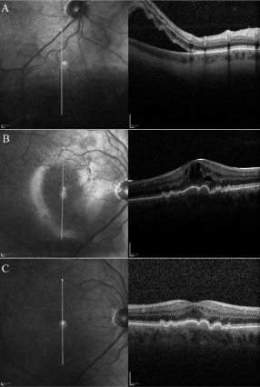
Figure 1: (A) OCT of the right eye1 day prior to silicone oil placement showing sub-retinal fluid with an inferior retinal detachment. (B) OCT reveals CME 1 month after silicone oil placement. (C) Resolution of CME1 month following silicone oil removal.
Case 2
A 53-year-old man presented with a 2-month history of worsening vision in his left eye. He had undergone pars plana vitrectomy with silicone oil tamponade 9 months prior for a macula-sparing rhegmatogenous retinal detachment OS. Silicone oil was removed 4 months after vitrectomy with an uneventful phacoemulsification and posterior chamber intraocular lens implant. On examination, his BCVA were 20/25 OD and 20/80 OS, fundoscopy revealed drusen and drusenoid pigment epithelial detachments without SRF or CME (Figure 2A). The patient developed a recurrent retinal detachment and subsequently underwent pars pana vitrectomy with silicone oil tamponade. At POM 1, BCVA was 20/400 OSCME (Figure 2B). Silicone oil was removed 4.5 months later. At POM1 month, BCVA improved to 20/70 OS with complete resolution of CME (Figure 2C).
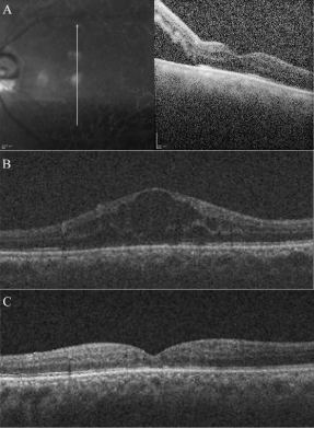
Figure 2: (A) OCT of the left eye showing sub-retinal fluid inferior to fovea from recurrent macula-involving rhegmatogenous retinal detachment. (B) OCT showing CME 1 month after silicone oil placement. (C) Resolution of CME 1 month following silicone oil removal.
Case 3
A 61-year-old man underwent surgical repair of a maculainvolving rhegmatogenous retinal detachment with pars pars plana vitrectomy with pneumaticretinopexy OD with hand motion vision under gas. OCT prior to surgery revealed a flat macula without SRF (Figure 3A). At post-operative week (POW) 1, the patient developed a recurrent retinal detachment with proliferative vitreoretinopathy and vitreous hemorrhage. The patient emergently underwent pars pars plana vitrectomy with silicone oil tamponade, phacoemulsification and posterior chamber intraocular lens implant OD. At POW 1, BCVA was 20/100 OD. Fundoscopy revealed a flat macula without CME. At POM 1, BCVA was 20/100 OD, and OCT showed new CME OD (Figure 3B). Silicone oil was removed 3.5 months later. At POM 1, BCVA improved to 20/60 OD, and fundoscopy revealed attached retina with a flat macular and resolution of CME (Figure 3C).
Figure 3: (A) OCT of the right eye showing a flat macula prior to maculasparking retinal detachment repair. (B) OCT showing CME 1 month after silicone oil placement. (C) Resolution of CME 1 month following silicone oil removal.
Discussion
Silicone oil with 5000 centistokes consistency was the preferred agent for tamponade in the reported cases due to the detachment location, the ability to control tamponade duration, and surgical preference in the treatment of concurrent proliferative vitreoretinopathy. The cases differed considerably with regards to patient age at presentation, baseline ocular health, clinical presentation, detachment severity, detachment configuration and extent of recovery and likely cause of retinal detachment. All three cases shared the clinical course of developing OCT-proven CME within 1 month after silicone oil placement, whileshowing visual acuityimprovement and CME resolution within 1 month after silicone oil removal.
CME formation has been linked to ocular inflammation caused by traumatic insults in procedures like cryotherapy, scleral buckle, and phacoemulsification during cataract extraction [4]. Given the temporal relationship of CME formation and silicone oil placement, traumatic insults caused by vitrectomy or endophotocoagulation is unlikely to cause CME in our patients. Silicone oil injection has been reported to stimulate an inflammatory reaction in clinical and experimental studies, though this response has never been shown to cause CME formation [5]. Interestingly, the precipitous appearance and resolution of CME within 1 month of silicone oil exposure and withdrawal coincides with Wichham et al. findings that silicone oil placement triggers a macrophage-mediated inflammatory reaction that presists for up to one month following silicone oil removal [6]. Silicone oil may also exacerbate intraocular inflammation by impairing the potassium ion buffering function of Müeller cells, which may lead to intra-retinal excitotoxicity and neuronal damage [7,8]. The exact nature of the pathology in our patients is unclear, but the resolution of CME following silicone oil removal may represent removal of the inflammatory nidus and subsequent recovery.
Tractional forces have also been implicated in CME formation [9]. Our incomplete understanding of the adhesive forces of silicone oil was first realized when silicone oil tamponade was utilized in patients with silicone intraocular lens. Experimental studies since then have demonstrated silicone oil has varying adhesive interactions with different materials and surfaces [10]. If non-uniform interactions are present between the silicone oil tamponade and the retinal surface, the resultant tractional forces may cause CME analogous to the anterior vitreomacular traction caused by Posterior Vitreous Detachment (PVD) [9]. If this hypothesis is correct, the abrupt resolution of CME following silicone oil removal correlates well with the resolution of foveal thickening observed with relief of vitreomacular traction in PVD [9].
Inflammatory and mechanical forces may play a role in CME development with use of silicone oil tamponade. Since most cases of CME resolve spontaneously, the decision to remove a silicone oil tamponade to expedite visual acuity improvement should be weighed against the adverse effects associated with premature tamponade removal.
References
- Abrams GW, Azen SP, McCuen BW 2nd, Flynn HW Jr, Lai MY, Ryan SJ. Vitrectomy with silicone oil or long-acting gas in eyes with severe proliferative vitreoretinopathy: results of additional and long-term follow-up. Silicone Study report 11. Arch Ophthalmol. 1997; 115: 335-344.
- Schwartz SG, Flynn HW Jr, Lee WH, Wang X. Tamponade in surgery for retinal detachment associated with proliferative vitreoretinopathy. The Cochrane database of systematic reviews. 2014; 2: CD006126.
- Falkner CI, Binder S, Kruger A. Outcome after silicone oil removal. Br J Ophthalmol. 2001; 85: 1324-1327.
- Valldeperas X, Romano MR, Wong D. Resolution of cystoid macular oedema after retinal detachment repair: is intravitreal triamcinolone useful? Eye. 2006; 20: 1321-1322.
- Sparrow JR, Matthews GP, Iwamoto T, Ross R, Gershbein A, Chang S. Retinal tolerance to intravitreal perfluoroethylcyclohexane liquid in the rabbit. Retina. 1993; 13: 56-62.
- Wickham LJ, Asaria RH, Alexander R, Luthert P, Charteris DG. Immunopathology of intraocular silicone oil: retina and epiretinal membranes. Br J Ophthalmol. 2007; 91: 258-262.
- Cazabon S, Groenewald C, Pearce IA, Wong D. Visual loss following removal of intraocular silicone oil. Br J Ophthalmol. 2005; 89: 799-802.
- Winter M, Eberhardt W, Scholz C, Reichenbach A. Failure of potassium siphoning by Muller cells: a new hypothesis of perfluorocarbon liquid-induced retinopathy. Investigative ophthalmology & visual science. 2000; 41: 256-261.
- Johnson MW. Tractional cystoid macular edema: a subtle variant of the vitreomacular traction syndrome. Am J Ophthalmol. 2005; 140: 184-192.
- Apple DJ, Isaacs RT, Kent DG, Martinez LM, Kim S, Thomas SG, et al. Silicone oil adhesion to intraocular lenses: an experimental study comparing various biomaterials. Journal of cataract and refractive surgery. 1997; 23: 536-544.