Perspective
Austin J Clin Ophthalmol. 2024; 11(6): 1198.
Adult Versus Pediatric Tomogram - A Perspective on Optical Coherence Tomography (OCT)
SR Dasari*
Consultant Ophthalmologist, Comprehensive, Member of AAO, India
*Corresponding author: SR Dasari, Consultant Ophthalmologist, Comprehensive, Member of AAO, India. Email: sdasari240@gmail.com
Received: November 12, 2024; Accepted: December 04, 2024; Published: December 11, 2024
Perspective
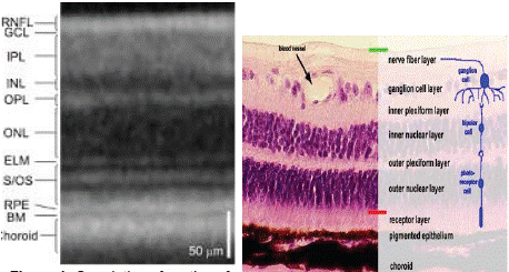
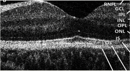
Optical Coherence Tomography (OCT) has been utilised to diagnose, animate and initiate thoroughly, examination of the retina. This form of noninvasive procedure in the field of Pediatric Ophthalmology, be it handheld, speckle, doppler or split using various domains like time (TD-OCT), spectral (SD-OCT) or Enhanced Depth Imaging (EDI-OCT) or Swept Source (SS-OCT), or Split Spectrum Amplitude (SSADA) optical coherence tomography has revolutionised the field of ophthalmology. Many a obvious pathology in the field of Pediatric Ophthalmology from retinal hyperplasia, congenital retinoschisis, retinal edema in retinopathy of prematurity to optic nerve disc ratios in optic nerve hypoplasia and congenital glaucoma have been visualised. Here we illustrate a simple picture of OCT in adult versus pediatric highlighting the areas of retina from inner retina to outer retina with respect to OCT and correlating it with histology of the ten layers of the adult retina. We further address the above narrative of how the optical coherence tomography has further enhanced the area by showing a correlation of adult OCT with pediatric OCT which has been adjusted for gestation age and neonates. All reports have shown 1) migration of inner retinal layers (Ganglion cell layer, inner plexiform layer, inner nuclear layer and outerplexiform layer) away from the central fovea. 2) migration of cone photoreceptors into central fovea. 3) elongation of outerretinal layers (outer nuclear layer, inner segment and outersegment IS/OS) with increasing age 1 thus explaining the presence of inner retinal layers and absence of cones in central fovea in neonates. Whereas inner retinal layers are absent and cone photoreceptors are present in central fovea on adult OCT. As the ten layers of the retina have been named, so also other minutia of histological relevance such as the External Limiting Membrane (ELM), the Myoid zone and the Photoreceptor Integrity Line (PIL) also called the ellipsoid zone at the IS/OS junction, the Interdigitation Zone (IZ) above the RPE followed by the Bruchs membrane have been identified in tomograms. Inner Segment (IS) or inner zone and Outer Segment (OS) or outer zone of photoreceptor layer have been nomenclatured in all tomograms. An ellipsoid zone is between external limiting membrane and inner photoreceptor segment. So also, a zone called myoid zone which is a lucency above the ellipsoid zone. The presence of Interdigitation Zone (IZ) is thought to be marker for retinal maturity. The Interdigitation Zone (IZ) is the last band to mature and is almost never visualised prior to 46-47 weeks gestation age. It is often absent on tomograms taken from premature and younger term infants 1 . Retinal Nerve Fibre Layer thickness (RNFL) axons which account for about 700,000 to 1.4 million, decrease at a rate of 0.75% year thus resulting in decrease in thickness of RNFL with age 2 . Optic disc diameters vary with patients age, sex and race. Optic disc measurements with the constituent optic cup and neuroretinal area vary with axial length, refractive error and show ratios which have consistent measurements irrespective of the methods of procurring the data 3.
    Location
    Average
    SD
    Max
    Min


    Superior
    136.5
    16.2
    168
    98


    Nasal
    87.9
    22.3
    135
    147


    Inferior
    141.5
    17.2
    176
    118


    Temporal
    69.8
    12.2
    103
    54
Table 1: The thickness of the pediatric retina (Retinal Nerve Fibre Layer
Thickness)1 in μm.
    Location
    Average
    +/-SD


    Superior
    124.2
    17.9


    Nasal
    80.9
    18.1


    Inferior
    126.1
    17.8


    Temporal
    69
    12.7
Table 2: The thickness of adult retina or retinal nerve fibre layer thickness
(RNFL)2i in um.
    Dimensions
    Average


    Horizontal
    1.53 mm


    Vertical
    1.79 mm


    Disc area
    2.2 mm2


    Optic cup
    0.7


    Cup: Disc horizontal
    0.46


    Vertical
    0.48
Table 3: Dimensions and Optic disc ratios of Pediatric optic disc.
    Dimensions
    Average
    Min
    Max


    Horizontal
    1.77 mm
    1.2
    1.7


    Vertical
    1.88 mm
    1.87
    1.96


    Disc area
    2.35 mm2
    1.84
    2.35 mm2


    Optic cup
    1.04
    1.04
    0.89


    Cup: Disc Horizontal
    0.48


    Vertical
    0.46
Table 4: Dimensions and Optic disc ratios of adult optic disc.

    Figure 1: Correlation of section of retina on OCT versus retinal histology
specimen.

Figure 2: Pediatíic retinal tomogram (OCL).
References
- Helena Lee, Frank A Proudlock, Irene Gottlob. Pediatric Optical Coherence Tomography in Clinical Practice—Recent Progress. Invest Ophthalmol Vis Sci. 2016; 57: OCL69–OCL79.
- Determinants of Normal Retinal Nerve fibre Layer Thickness measured by Stratus OCL- Ophthalmology. 2007; 114: 1046-1052.
- Donald Budenz, Douglas Anderson, Rohit Varma, Joel schuman, Louis Cantor, Jonathan Savell, et al. Optic disc rim area is related to disc size in normal subjects. Arch of opthalmology. 1987; 105: 1683-1685.