
Case Report
Austin J Clin Ophthalmol. 2023; 10(7): 1166.
Toxocara Optic Neuropathy: About 02 Cases
Hasnaoui Ihssan*; Amine Krichen; Salma Hassina; Bardi Chaimaa; Louai Serghini; Abdellah Elhassan
Department of Ophtalmologie B & Faculty of Medicine and Pharmacy, Hospital of Specialities, University Mohamed V, CHU ibn Sina Rabat, Morocco
*Corresponding author: Hasnaoui I Department of Ophtalmology B, Faculty of Medicine and Pharmacy, University Mohamed V, Av. Abderrahim Bouabid, 10100 Rabat, Morocco. Email: ihssanhasnaoui@gmail.com
Received: September 07, 2023 Accepted: October 14, 2023 Published: October 21, 2023
Abstract
Ocular toxocariasis is recognized to be a cause of childhood blindness. It usually results as a sequel of systemic infestation with the second or third stage larva of T. cani (visceral larval migrans). It is an uncommon cause of uveitis that mainly affects younger patients. Inflammation is typically unilateral and presents as either a granuloma in the peripheral or posterior retina or a moderate to severe vitreous inflammation mimicking endophthalmitis. We report two cases of children with ocular toxocariasis with posterior granuloma, negative for specific serodiagnosis. An anterior chamber puncture of the anterior chamber and an ELISA test with homologous antigens of Toxocara canis on the acqueous humor made the diagnosis diagnosis.
Keywords: Optic neuropathy; Toxocariasis; Ocular toxocariasis; Toxocara optic neuropathy
Introduction
In many parts of the world, parasitic infections of the eye are a major cause of blindness [1].
Ocular Toxocariasis (OT) is an uncommon disease that occurs primarily in young patients. It affects females and males with approximately equal frequency. Toxocariasis or visceral larva migrans is a cosmopolitan parasitic zoonosis caused by Toxocara canis or Toxocara cati or, even more rarely, by nematode larvae from wild animals. The prevalence rate in industrialized countries is 2-5% in urban areas, 37% in rural areas. Major forms include visceral larva migrans syndrome, neurological toxocariasis and ocular toxocariasis. Ocular involvement is rare, and manifests itself essentially in three forms: pan-uveitis, posterior granuloma and peripheral granuloma.
Patients and Observation
Case 1
A 10-year-old child with no medical history was admitted for a sudden vision loss of the right eye. On ophthalmological examination, the visual acuity was reduced to hand motion in the right eye, the anterior segment was calm; the fundus showed intermediate uveitis and a parapapillary granuloma surrounded by retinal elevation (Figure A); the ophthalmological examination of the left eye did not reveal any abnormality. Ocular ultrasound revealed a small, heterogeneous echogenic formation forming a voluminous papillary protrusion with no visible calcification (Figure B). Biological tests revealed a hypereosinophilia of 920/mm3, a normal chest X-ray and a negative tuberculin skin test. Blood serologies were negative for both toxoplasmosis and toxocariasis (Toxocara canis ELISA technique). On the aqueoushumor, the PCR test for toxoplasmosis DNA was negative, and the ELISA immunodiagnosis for toxocariasis was positive at 1.725. Western blot also confirmed the diagnosis. Appropriate treatment was instituted, including general corticosteroid therapy (prednisone 1mg/kg/d) and minute antiparasitic treatment with albendazole. At the end of the treatment, the uveitis disappeared without any improvement in visual acuity.
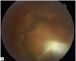
Figure A: Yellowish-white parapapillary focus with sharp borders surrounded by tractional retinal elevation.
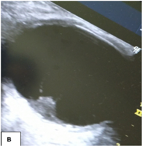
Figure B: A solid, highly reflective papillary mass with no calcification.
Case 2
A 6-year-old child was admitted for a progressive vision loss with esotropia of the left eye. He had no previous history of esotropia, or any similar case in the family. Clinical examination on admission showed visual acuity of 10/10 in the right eye, and hand motion in the left eye. Slit-lamp and fundus examinations revealed no abnormalities in the right eye. The left eye had a clear cornea, a good anterior chamber and no synechiae. The fundus showed hyalitis ++, a yellowish-white parapapillary granuloma in the shape of a glove finger, with no flange, active focus or scarring (Figure C).
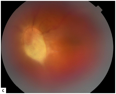
Figure C: Yellowish-white parapapillary granuloma in a 6-year-old boy with Toxocara canis infection.
The chest X-ray was normal and the tuberculin intradermal test negative. Blood serologies were negative for both toxoplasmosis (IgG and IgM) and toxocariasis. On the aqueous humor sample, toxoplasmosis DNA polymerase chain reaction was negative, and toxocariasis immunodiagnosis by ELISA was positive at 1.701. The Western blot also confirmed this positivity. The child received general corticosteroid therapy combined with albendazole at a dose of 400mg/day for 15 days, with progressive reduction of the corticosteroid over 6 weeks. The evolution was marked by rapid regression of the hyalitis, at the end of the treatment, the uveitis disappeared but the prognosis was poor given the location of the granuloma.
Discussion
Ocular toxocariasis is more common in developing countries and in non-urbanized areas. It tends to affect young children through ingestion of embryonated eggs, and accounts for 1-2% of uveitis. The disease is unilateral in most cases, with mild to moderate intermediate or diffuse inflammation. Clinical presentation varies: one half of all eyes have a granuloma in the peripheral retina, one quarter has a granuloma in the macula, and one quarter has endophthalmitis. A granuloma can also be present on the optic nerve. The most common clinical signs are vitreous inflammation, cystoid macular edema, and vitreoretinal traction strands leading to the optic disk and/or a granuloma. The diagnosis of OT is difficult and in the majority of cases remains only presumptive. Standard diagnostic methods for ocular Toxocara are funduscopy, imaging, and serologic testing for detection of IgG-specific antibodies in serum, aqueous humor or vitreous (Elisa technique) [7]. Negative serology does not rule out the diagnosis; in this 2 cases, serum ELISA results were positive for both patients, in both patients, visual acuity is diminished at presentation. The common causes of vision loss are vitreous inflammation and traction retinal detachment.
Imaging techniques can help with the diagnosis when the fundus is inaccessible, by looking for complications such as tractional retinal detachment, and above all to eliminate the hypothesis of retinoblastoma, given the seriousness of this pathology in children. Treatment is not systematic, depending on the degree of inflammation and location of the granuloma on the fundus, and relies on systemic corticosteroid therapy combined with anti-parasitic agents (Thiabendazole or Albendazole) [8].
Conclusion
Toxocariasis is a widespread zoonosis with a poor prognosis; Our work illustrates the seriousness of toxocariasis when it affects the eye. In all cases of uveitis, toxocariasis serology by enzyme-linked immunosorbent assay or Western Blot technique should be requested in order to institute urgent treatment.
References
- Cunningham ET Jr. Uveitis in children. Ocular Immunol Inflamm. 2000; 8: 251–261.
- Darrell RW, Wagener HP, Kurland LT. Epidemiology of uveitis: incidence and prevalence in a small urban community. Arch Ophthalmol. 1962; 68: 502–514.
- Vadot E, Barth E, Billet P. Epidemiology of uveitis: preliminary results of a prospective study in Savoy. In: Saari KM, ed. Uveitis Update. Amsterdam: Elsevier Science Publishers; 1984; 13.
- Perkins ES, Folk J. Uveitis in London and Iowa. Ophthalmologica. 1984; 189: 36–40.
- Henderly DE, Genstler AJ, Smith RE, Rao NA. Changing pattern of uveitis. Am J Ophthalmol 1987; 103: 131-136.
- Rodriguez A, Calonge M, Pedroza-Seres M, et al. Referral patterns of uveitis in a tertiary eye care center. Arch Ophthalmol. 1996; 114: 593–599.
- do Lago A, Andrade R, Muccioli C, Belfort R. Optical coherence tomography in presumed subretinal Toxocara granuloma: case report. Arq Bras Oftalmol. 2006; 69: 403-405.
- Shields JA. Ocular toxocariasis: a review. Surv Ophthalmol. 1984; 28: 361–381.