
Case Report
Austin J Clin Ophthalmol. 2023; 10(4): 1152.
Central Retinal Artery Occlusion with Sparing of a Double Cilioretinal Artery: A Case Report
Najoua El Moubarik*; Hamza Lazaar; Kenza Tazi; Saad Bnechekroun; Nourdine Boutimzine; Abdellah Amazouzi; Samira Tachfoufi; lalla Ouafae Cherkaoui
Department of Ophthalmology “A”, Ibn Sina University Hospital (Hôpital des Spécialités), Mohammed V University, Morocco
*Corresponding author: Najoua El Moubarik Department of Ophthalmology “A”, Ibn Sina University Hospital (Hôpital des Spécialités), Mohammed V University, Rabat, Morocco. Email: najoua.elmoubarik@gmail.com
Received: February 27, 2023 Accepted: April 05, 2023 Published: April 12, 2023
Abstract
Central Retinal Artery Occlusion (CRAO) is a rare condition that is functionally life-threatening. It is the equivalent of an ischemic stroke.
The cilio retinal artery is an anatomical variant that allows supplementing the vascularization of the macula in case of occlusion of the central retinal artery.
CRAO consists of the following four distinct clinical entities: non-arteritic CRAO (NA-CRAO), transient NA-CRAO, NA-CRAO with cilioretinal artery sparing, and arteritic CRAO. Clinical characteristics, visual outcome, and management very much depend upon the type of CRAO.
We report the case of a 29-year-old patient, with a history of recurrent angina, who presented with a sudden decrease in visual acuity in his left eye two days before his consultation. The diagnosis of central retinal artery occlusion sparing a double cilioretinal artery was evoked and treated early, allowing a significant visual recovery.
Keywords: CRAO; Cilio retinal artery; Embolic etiology; Urgent management; Functionally prognostic
Introduction
Central Retinal Artery Occlusion (CRAO) is a rare condition that is functionally prognostic.
The cilio retinal artery is an anatomical variant present in 20% of the population; it allows supplementing the vascularisation of the macula in case of occlusion of the central retinal artery [1].
Case Report
We report the case of a 29-year-old patient, with a history of recurrent angina, who consulted for a drop in visual acuity in the left eye that occurred suddenly two days before his consultation.
The best visual acuity was 4/10 in the left eye.
The fundus examination revealed an obliteration of the central artery of the left retina with a whitish ischemic retinaledema sparing two horizontal territories: one at the level of the interpapillomacular and macular bundle and the other nasal to the papilla measuring two papillary diameters (Figure 1). The examination of the adelphic eye was normal.
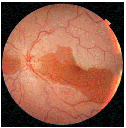
Figure 1: Retinophotography: Visualization of retinal edema sparing both territories of the cilioretinal arteries.
Fluorescein angiography showed a prolongation of the retinal arm time, a filling defect of the 4 branches of the central retinal artery with perfusion of two cilio retinal arteries (Figure 2).
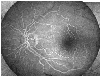
Figure 2: Fluorescein angiography: Visualization of a prolonged retinal arm time, a filling defect of the 4 branches of the central retinal artery with perfusion of two cilio retinal arteries.
The OCT showed the presence of a diffuse retinal thickening sparing the retina dependent on the two cilio retinal arteries with a hyperreflexia of the inner retinal layers (Figure 3).
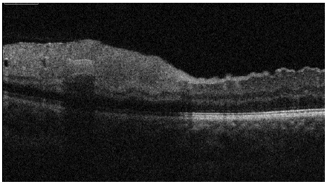
Figure 3: Macular OCT showing diffuse retinal thickening sparing the retina dependent on the two cilio retinal arteries with hyperreflectivity of the inner retinal layers.
The etiological work-up revealed the presence of an aortic narrowing with an intra-cavity thrombus requiring hospitalization in an intensive care unit for anticoagulation.
The evolution was marked by an improvement of the visual acuity to 7/10, with a decrease of the retinal thickening (Figure 4).
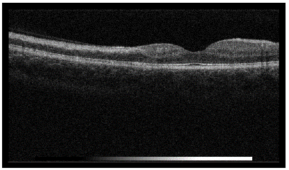
Figure 4: OCT showing the decrease of retinal thickening.
Discussion
CRAO is a severe vascular event equivalent to an ischemic stroke. It usually occurs in adults over 40 years of age with cardiovascular risk factors. Its incidence is about 1/10,000/year [1].
The retinal arterial vascularisation is of the terminal type. Any occlusion of the central retinal artery or its branches leads to irreversible downstream ischemia of the inner retinal layers [2]. In about 20% of cases, there are cilioretinal arteries involved in the vascularisation of the macular region. They originate from the posterior ciliary circulation and are therefore spared during RCA occlusion. These patients with a cilioretinal artery often retain good central visual acuity because the vascularisation of the macula is intact,
The particularity in our case was the presence of two cilioretinal arteries allowing the sparing of two retinal territories on both sides of the papilla, and thus the conservation of visual acuity.
Few cases of CRAO with sparing of double cilio retinal artery have been reported in the literature.
Four mechanisms can be distinguished in the occurrence of CRAO: embolism of carotid or cardiac origin is the most frequent in practice, arterial etiology (rarer 5% of CRAO), and local compressive cause and coagulation disorders [2].
The classic picture associates a cherry red macula, a granular current in the vessels. Ischemic edema of the retinal ganglion cells results in a milky white appearance of the retina. When a cilioretinal artery exists, its vascularisation territory remains normally colored, which contrasts with the rest of the whitish ischemic retina [4].
The emergency ophthalmic workup should include fluorescein angiography to show the prolonged retinal arm time, delayed choroidal, arterial and arteriovenous filling. Macular optical coherence tomography imaging is not necessary for diagnosis but provides information on the retinal status.
The ophthalmological workup should not delay the general workup, which includes a clinical cardiovascular and neurological examination associated with an electrocardiogram in the ophthalmological emergency room, before being transferred to an appropriate department to continue the etiological workup (ultrasound of the supra-aortic trunks, cardiac ultrasound, MRI) [3].
Conclusion
CRAO should be managed as an acute stroke, with the objective of early reperfusion and secondary prevention. Collaboration between ophthalmologists and vascular neurologists is essential.
References
- Hayreh SS. Acute retinal arterial occlusive disorders, Prog Retin Eye Res. 2011; 30: 359-394.
- P Lallemand, D Gaucher. L’OACR en pratique: quel bilan, quel prognostic, quel traitement, réalités ophtalmologiques # 201_Mars 2013_Cahier 1.
- Mehta N, Marco RD, Goldhardt R, Modi Y. Central Retinal Artery Occlusion: Acute Management and Treatment. Curr Ophthalmol Rep. 2017; 5: 149-159.
- Wolff B. Occlusions artérielles rétiniennes. Encyclopédie médico-chirurgicale. 2012.