Clinical Image
Austin J Clin Ophthalmol. 2023; 10(3): 1146.
Multimodal Retinal Imaging of Paediatric Focal Choroidal Excavations
Clarke K*, Mahgoubn K, Hurtikova K and Thomas D
Moorfields Eye Hospital, NHS Foundation Trust, UK
*Corresponding author: Clarke KMoorfields Eye Hospital, NHS Foundation Trust, 162 City Rd, London EC1V 2PD, UK
Received: February 13, 2023; Accepted: March 14, 2023; Published: March 21, 2023
Clinical Image
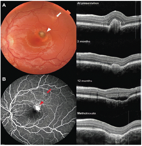
A 13-year-old female presented with reduced visual acuity of 6/36. Fundal imaging revealed foveal and eccentric focal choroidal excavations (Figure 1A). Optical coherence tomography confirmed the presence of two cavitating deformations of the choroid with disturbance of the foveal outer retinal layers and corresponding subretinal haemorrhage [1]. The foveal lesion was associated with a choroidal neovascular membrane and a suspected inflammatory element. Late-phase fluorescein angiography (Figure 1B) revealed large central leakage (red arrowhead) and faint hyperfluorescence overlying the eccentric lesion (red arrow).

Figure 1: A) Colour fundus photography of the left eye showing both the foveal (white arrowhead) and eccentric (white arrow) focal choroidal excavations. B). Late-phase Fundus Fluorescein Angiography (FFA) of left showing large central leakage (red arrowhead) and faint hyperfluorescence overlying the eccentric lesion (red arrow). Serial Optical Coherence Tomography (OCT) images of central FCE lesion show it’s relapsing and remitting course and resolution upon commencing methotrexate.
Two months after initiating treatment with intravitreal bevacizumab and prednisolone, the patient’s vision improved to 6/6. Over the next two years, the foveal lesion took a relapsing and remitting course, where at times, the retina conformed to the shape of the choroid, and at others, it was non-conforming due to subretinal fluid [2]. Both excavations have remained stable for one year following the initiation of methotrexate.
Acknowledgement
We would like to thank the patient whose images are used in this article for contributing to ophthalmic research.
Conflicts of Interest/Competing Interests
The authors declare no conflicts of interest.
Consent to Participate
The patient has given verbal and written consent for use of their clinical imaging in this publication Consent for publication (According to ICMJE Recommendations for protection of research participants)
References
- Park KA, Oh SY. The absence of focal choroidal excavation in children and adolescents without retinal or choroidal disorders or ocular trauma. Eye. 2015; 29: 841-2.
- Margolis R, Mukkamala SK, Jampol LM, Spaide RF, Ober MD, et al. The expanded spectrum of focal choroidal excavation. Archives of ophthalmology. 2011; 129: 1320-5.