
Research Article
Austin J Clin Ophthalmol. 2014;1(7): 1035.
Surgical Management of Congenital and Aponeurotic Ptosis – Cosmetic Outcome
Rizvi SAR*, Yousuf S and Gupta Y
Institute of Ophthalmology, Jawaharlal Nehru Medical College, India
*Corresponding author: Rizvi SAR Ophthalmic Plastic Surgery and Ocular Oncology Services, Institute of Ophthalmology, Jawaharlal Nehru Medical College, AMU, Aligarh, UP, 202002. India
Received: August 20, 2014; Accepted: September 18, 2014; Published: September 20, 2014
Abstract
To determine the cosmetic results following the surgical correction of congenital and acquired ptosis. A retrospective review of the surgical outcome of 64 eyes of 57 cases of congenital and acquired ptosis operated by single surgeon between January 2009 and December 2013 was evaluated. 21 cases (23 eyes) underwent a levator resection procedure, 23 cases (28 eyes) were operated by frontalis suspension using silicone rod, 6 cases (9 eyes) were operated by levator advancement procedure for aponeurotic ptosis and 4 cases were corrected by fasanella servant surgery and 3 cases by mullerectomy. Cosmetic outcome were considered to be good if post operative symmetry between two eyelids is ≤1mm,fair if symmetry is between 1.5 -2.0 mm and poor if it is >2mm at 6 weeks of follow up. Out of total 57 cases, a good cosmetic result was achieved in 45 cases (79%), fair in 7 cases (12%) and poor result was seen in 5 cases (9%). An acceptable cosmetic result was achieved in 91% of all cases. In 9% of cases with poor cosmesis, re surgery was required. Regular follow-ups will help in adequate management of complications and recurrence of ptosis.
Keywords: Congenital; Acquired; Ptosis; Surgery; Cosmesis
Introduction
Ptosis is defined as an abnormal low lying upper eyelid margin with eyes in primary gaze [1]. Ptosis is considered congenital if present at birth or within the first year of life [2]. The most common cause of congenital ptosis is localized myogenic dystrophy of levator palpebrae superioris. Few cases of congenital ptosis result from genetic or chromosomal defects or neurological dysfunction [3]. Simple congenital ptosis with normal superior rectus function is seen in 95% cases. Congenital ptosis can be associated with blepharophimosis syndrome (5%) and synkinetic ptosis (5%) which includes Marcus Gunn jaw winking ptosis and misdirected third nerve ptosis. In patients with congenital ptosis reduced levator function, absent lid crease and lid lag are characteristic findings. Acquired ptosis is classified into Neurogenic, Myogenic, Aponeurotic and Mechanical Ptosis. Majority of cases are due to aponeurotic causes such as involutional changes, disinsertion or dehiscence. In aponeurotic cases, the lid crease is higher due to disinsertion of the posterior aponeurotic attachment to the tarsus.
Material and Methods
A retrospective analysis was made of all 64 eyes (57 cases) of surgically corrected congenital ptosis operated by single surgeon between January 2009- December 2013. Cosmetic outcome was determined by post operative eyelid symmetry at 6 weeks of follow-up. It was graded as good if ≤1mm; fair if between 1.5 -2.0 mm and poor if >2mm.
Surgical procedures
Fifty-one cases were operated for congenital ptosis by four surgical techniques. The mean follow-up period was six weeks. Twenty-one patients underwent levator resection procedure. Two patients had bilateral procedures for bilateral ptosis giving a total of twenty-three procedures. Twenty-three patients underwent frontalis suspension using silicone rod. Five of the patients had bilateral procedures for bilateral ptosis giving a total of twenty-eight procedures. Four patients underwent Fasanella Servat procedure and three patients underwent mullerectomy. Six cases were operated for aponeurotic ptosis with levator advancement procedure.
Results
In all the 51 patients of congenital ptosis, ptosis was recognised since birth. All the patients had decreased levator function, absent lid crease and lid lag in down gaze.
In 21 cases of levator resection, 16(76%) cases (1 bilateral) achieved good cosmetic results (Figure 1a,1b), 3(14%) cases (1 bilateral) showed fair result and 2 (10%) cases had poor surgical outcome. The two patients (2 eyes) who showed poor cosmetic result were young children and poor result may be attributed to improper preoperative evaluation. (Figure 2).
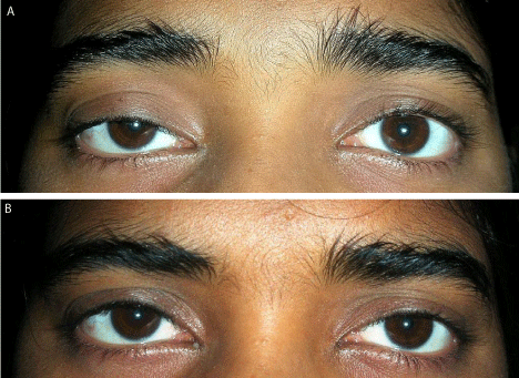
Figure 1a,b : Showing Preoperative and Postoperative Results of Levator Resection.
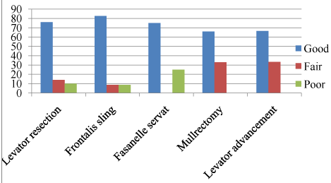
Figure 2 : Showing Cosmetic Outcome of Ptosis Surgery.
Out of 23 cases of frontalis suspension, 19(82.6%) cases ( 4 bilateral) shows good cosmesis (Figure 3a,3b), 2(8.6%) cases (1 bilateral) had fair results while 2 (8.6%) cases showed poor results.1case failed due to granuloma formation and subsequent silicone rod extrusion and 1 case had recurrence at 6 weeks. (Figure 2).
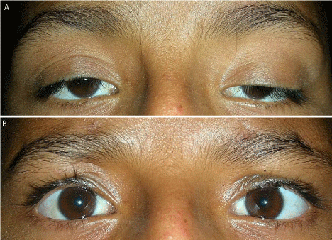
Figure 3a,b : Showing Preoperative and Postoperative Results of TarsoFrontalis Sling Surgery.
Fasanella servat surgery was done in 4 cases, out of which 3(75%) cases had good cosmetic results (Figure 4a,4b), while 1(25%) case had poor cosmesis. Lately, mullerectomy was planned in 3 cases of mild ptosis out of which 2(66.66%) cases showed good result (Figure 5a,5b), and 1(33.33%) case had fair result (Figure 2 ).
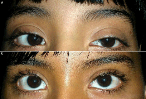
Figure 4a,b : Showing Preoperative and Postoperative Results of Fasanella Servat Surgery.
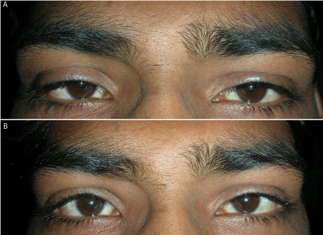
Figure 5a,b : Showing Preoperative and Postoperative Results of Mullerectomy.
In 6 cases of aponeurotic ptosis, levator advancement surgery was done. 4 cases (66.6%) showed good cosmetic result (Figure 6a,6b) while 2 cases (33.3%) showed fair results (Figure 2).
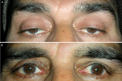
Figure 6a,b : Showing Preoperative and Postoperative Results of Levator Advancement Surgery.
Discussion
The choice of operative method for congenital ptosis depends on the age of patient, degree of ptosis, function of the levator muscle and the status of cornea. The accepted techniques for surgical repair of congenital ptosis are Fasanella-Servat procedure, Mullerectomy, levator aponeurosis resection and brow frontalis suspension. The determining factors as to which procedure is performed are the severity of ptosis and the amount of levator function. Levator function is the most important eyelid measurement in terms of surgical planning as the effectiveness of certain surgical procedures rests solely on the integrity of levator muscle function.
Levator resection is employed for cases with levator function (≥4mm). The overall success rate in our series was 76% with poor results in 10.2% cases. The patients with poor results were children and poor results could be due to improper evaluation of levator function. Cates and Tyers reported a success rate of 75% in 100 patients with congenital ptosis who underwent levator resection [4]. In their study, the most common complication was under correction (19%).They found that the preoperative amount of levator function was the strongest predictor of a favourable outcome following levator resection surgery. Berlin and Vestal reported a 69% success rate in 58 cases of congenital ptosis [5]. Blomgren and Holmstrom published a 52% success rate in cases having a single operation in 55 eyelids [6]. In a report by Jordan and Anderson on 228 cases of levator resection, the success rate was 43% [7]. Most studies suggest that levator function and ptosis level before surgery are correlated with the outcomes of levator resection.
Frontalis suspension surgery is done in congenital ptosis with levator function <4mm. In our series of 23 patients (28 eyes) silicone sling was used for frontalis suspension. The success rate was 82.6%. Failure was seen due to rod extrusion and granuloma formation. Carter et al retrospectively studied 35 patients (61 eyelids) who underwent frontalis suspension using silicone sling [8]. Final lid height was good in all 100% eyelids. Revision was required in 7% cases, migration of sling occurred in 5% cases and extrusion of sling in 5% cases. Morris et al retrospectively studied 89 patients (110 eye lids) had silicone rod frontalis suspension surgery [9]. Satisfactory result was seen in 63% cases. Complications occurred in 11% cases.
Fasanella Servat Procedure or Mullerectomy is the procedure of choice in good levator function. In our series Fasanella Servat had success rate of 75% and Mullerectomy had success rate of 66.7%. Prokosch et al evaluated post operative success of 20 patients (23 eyelids) who underwent Fasanella Servat procedure [10]. Post operative eyelid symmetry was found in 90% of patients.
Levator advancement surgery is done for aponeurotic ptosis. In our series good results were seen in 67% of cases. Patel et al did a retrospective analysis of 71 eyelids that underwent levator advancement surgery and found a success rate of 87% [11].
In our series of 57 cases good cosmesis was seen in 45 cases (79%), fair cosmesis was seen in 7 cases (12%) and poor cosmesis in 5 cases (9%). In our series acceptable cosmetic result was seen in 91% cases. 9% cases had poor cosmesis and required re surgery. The conclusion drawn from this series are that in fair to good levator action levator resection is preferred form of surgery .When levator action is good surgery of choice is Fasanella servat or mullerectomy. In cases of poor levator action frontalis suspension surgery should be performed. The use of silicone rod resulted in a good cosmetic appearance. In aponeurotic ptosis levator advancement surgery is the preferred surgery. Regular follow-up is necessary for management of complications and recurrence.
References
- Beard C. Types of ptosis. Beard C, editors. In: Ptosis. 3rd edn. St. Louis: Mosby; 1981; 39-76.
- Sakol PJ, Mannor G, Massaro BM. Congenital and acquired blepharoptosis. Curr Opin Ophthalmol. 1999; 10: 335-339.
- Guercio JR, Martyn LJ. Congenital malformations of the eye and orbit. Otolaryngol Clin North Am. 2007; 40: 113-140.
- Cates CA, Tyers AG. Outcomes of anterior levator resection in congenital blepharoptosis. Eye (Lond). 2001; 15: 770-773.
- Berlin AJ, Vestal KP. Levator aponeurosis surgery. A retrospective review. Ophthalmology. 1989; 96: 1033-1036.
- Blomgren I, Holmström H. Anterior levator resection in congenital genuine blepharoptosis. A follow-up of 55 operated eyelids. Scand J Plast Reconstr Surg. 1986; 20: 189-195.
- Jordan DR, Anderson RL. The aponeurotic approach to congenital ptosis. Ophthalmic Surg. 1990; 21: 237-244.
- Carter SR, Meecham WJ, Seiff SR. Silicone frontalis slings for the correction of blepharoptosis: indications and efficacy. Ophthalmology. 1996; 103: 623-630.
- Morris CL, Buckley EG, Enyedi LB, Stinnett S, Freedman SE. Safety and efficacy of silicone rod frontalis suspension surgery for childhood ptosis repair. Journal Pediatric Ophthalmology. 2008; 45: 280-288.
- Prokosch V, Boehm MR, Choontanom R, Merté RL, Stupp T. Fasanella-Servat procedure for the surgical treatment of acquired ptosis of the upper eyelid. Klin Monbl Augenheilkd. 2011; 228: 29-34.
- Patel V, Salam A, Malhotra R. Posterior approach white line advancement ptosis repair: the evolving posterior approach to ptosis surgery. Br J Ophthalmol. 2010; 94: 1513-1518.