
Case Report
Austin J Clin Case Rep. 2025; 12(2): 1353.
Pleomorphic Adenoma of Upper Lip – A Rare and Unusual Presentation
Shah K, Pandhare K*, Landge J, Gavali P and Rahman A
Department of Oral and Maxillofacial surgery, Government Dental College and Hospital, Chh. Sambhaji Nagar, Maharashtra, India
*Corresponding author: Dr. Kshitija Pandhare, Department of Oral and Maxillofacial surgery, Government Dental College and Hospital, Chh. Sambhaji Nagar, Maharashtra, India Email: dhanakshitu.kp@gmail.com
Received: June 01, 2025 Accepted: June 19, 2025 Published: June 23, 2025
Abstract
Pleomorphic adenoma is most common benign salivary gland tumor. It is most commonly seen in parotid gland and salivary glands of palate but the occurrence of pleomorphic adenoma in rare in upper lip. This study aims to present a case of pleomorphic adenoma of upper lip in a 38 year old female. Radiograph and ultrasonography was done. Treatment was given as surgical excision of the tumor along with its capsule and clear margins are required due to risk of malignant transformation.
Introduction
The most frequent neoplasm of the salivary glands is pleomorphic adenoma (PA), also known as a benign mixed tumor [1]. It usually develops in the parotid gland but can also affect smaller salivary glands, like those in the upper lip [2]. Although Pleomorphic adenoma of the hard palate is very common, they are nevertheless extremely uncommon to appear in the upper lip.
Clinically the pleomorphic adenoma of upper lip is presented as a swelling that is firm and painless and appears similar to a traumatic fibroma or a mucocele. Because of the possibility of recurrence or malignant change, this tumor, although benign, require total surgical removal [3]. Because of clinical rarity and possible malignant potential precise diagnosis and treatment is required [4].
This study aims to present a case of pleomorphic adenoma of upper lip in a 38 year old female. Radiograph and ultrasonography were done. Treatment was given as surgical excision of the tumor along with its capsule and clear margins are required due to risk of malignant transformation.
Case Presentation
Patient Information
A 38 year old female patient came to Department of Oral and Maxillofacial surgery with a chief complaint of swelling in left side of upper lip. She was free of all comorbidities. Family history was negative.
Clinical Findings
Patient complaint of painless swelling present in upper lip since 6 months.
On examination, the swelling was firm, sessile, painless of size 1.2*0.8 cm. Swelling was slow growing and well defined. The mass had expanded outwards towards the skin rather than inwards towards the oral mucosa. Surrounding skin and mucosa appeared normal. No lymph node involvement.
Diagnostic Approach
Radiograph was done which did not show any changes.
Ultrasonography local was done which revealed 1.2*0.68 cm sized well defined hypoechoic lesion showing no vascularity on doppler study noted in upper lip suggestive of Epidermal inclusion cyst or a fibroma (Figure 1-6).
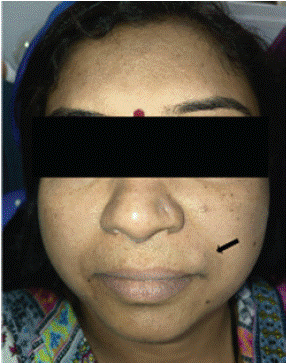
Figure 1: chromatogram of sample solution (80 μg/mL).
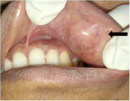
Figure 2: Swelling seen on left upper labial mucosa.
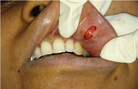
Figure 3: Incision given on mucosa.
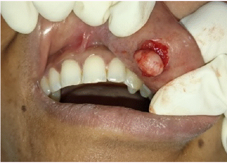
Figure 4: Blunt dissection done in order to keep the capsule intact.
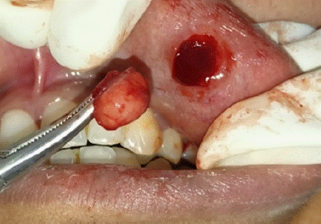
Figure 5: En-mass excision of lesion done.
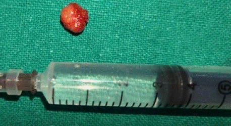
Figure 6: 1.2*0.68 cm lesion excised.
Treatment Approach
Under all aseptic precautions, under local anesthesia, an incision was given on the labial mucosa (via an intraoral approach) and blunt dissection was done. The lesion was excised in toto with capsule intact. Margins were cleared until we see underlying muscle. Suturing done with 3-0 Vicryl.
Final Diagnosis
The specimen was sent for Histopathological examination which showed H & E-stained section of soft tissue bits exhibits lesional tissue consisting tumor cells of glandular origin which are encapsulated by loosely arranged fibrous capsule. The tumor cells consist of epithelial and myoepithelial cells proliferating in the form of sheets and strands. Myoepithelial cells are showing varying morphology from spindle to rounded cells with eccentric nuclei and hyalinized eosinophilic cytoplasm giving plasmacytoid appearance. Duct like structures are lined by low cuboidal to columnar cells and lumen contain eosinophilic coagulum. Mucoid, myxoid areas are seen throughout the stroma and osseous areas are seen at places. Deeper section shows normal salivary gland acini and transverse and longitudinal sections of muscle fibre bundles (Figure 7,8).
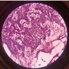
    Figure 7: H and E section showing myoepithelial cells showing eccentric
nuclei and hyalinized eosinophilic cytoplasm.
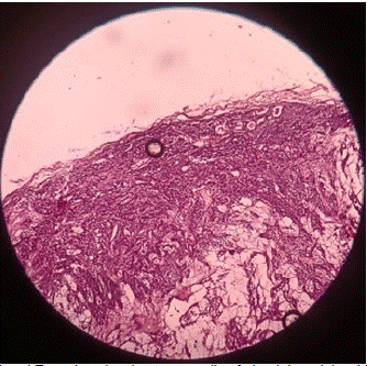
    Figure 8: H and E section showing tumor cells of glandular origin which are
encapsulated by loosely arranged fibrous capsule.
Overall features suggestive of "Pleomorphic adenoma".
Discussion
Pleomorphic adenoma is most common benign salivary gland tumor. It is most commonly seen in parotid gland and salivary glands of palate but the occurrence of pleomorphic adenoma in rare in upper lip. It has been hypothesized that the difference in the incidence of PA between the upper and lower lips can be ascribed to embryological development, as the upper lip is six times more likely to be afflicted than the lower [3]. There is complex fusion process in the upper lip. Three facial protuberances combine to make the upper lip which raises the risk of tumorigenesis in that area by increasing the possibility that ectopic embryonic cells that may become stuck. Also, the upper lip includes a large number of salivary glands than the lower lip. It is still unclear that what is the primary etiopathogenesis of PA. According to cytogenetic and molecular research, it has an epithelial origin with chromosome abnormalities at 8q12 and 12q15 [5,6].
It has a female predominance [7]. These tumors are most often diagnosed in the 4th and 6th decades of life. In the article given by Lotufo MA [5], pleomorphic adenoma of upper lip is seen in a 12 year old boy which suggests that it can be seen in any age. Bernier found that peak incidence of pleomorphic adenoma of upper lip is seen in age group of 30- 40 years with average of 33.2 years [8]. In our case patient was 38 year old female complaint of painless, slow growing swelling since 6 months. The prognosis is good if the tumor is excised in toto along with its capsule. It is most likely seen that benign lesions occur in upper lip where as malignant lesions in lower lip [9,10].
The tumor was excised en-mass with disease-free margins. Patient was kept for follow up of 6 months with no recurrence.
Conclusion
Pleomorphic adenoma has to be included in a differential diagnosis of the lesions appearing in upper lip with a history of slow growing and painless swelling. These lesions are to be considered for histopathological examination and are usually encapsulated so excisional biopsy with excision of capsule should be the treatment of choice.
References
- Shah BA, Singh AP, Sherwani AY, Ahmad SM. Pleomorphic adenoma of the upper lip: A rare case report. National Journal of Maxillofacial Surgery. 2020; 11: 289-291.
- Prakash C, Tanwar N, Dhokwal S, Devi A. A rare case report of pleomorphic adenoma of the upper lip: an unusual clinical presentation. National Journal of Maxillofacial Surgery. 2022; 13: S225-227.
- Ali RM, Ali RM, Qaradakhy AJ, Hiwa DS, Abdullah AM, Omar DA, et al. Pleomorphic adenoma of the lip: a case report and literature review. Journal of Surgical Case Reports. 2025; 2025: rjaf311.
- Mishra S, Pati N, Nayak AA. Pleomorphic Adenoma of Upper Lip: A Rare Case Report in a Tertiary Care Hospital. Asian Journal of Pharmaceutical and Health Sciences. 2017; 7.
- Lotufo MA, Júnior CA, de Mattos JP, França CM. Pleomorphic adenoma of the upper lip in a child. Journal of oral science. 2008; 50: 225-228.
- Debnath SC, Adhyapok AK. Pleomorphic adenoma (benign mixed tumour) of the minor salivary glands of the upper lip. Journal of maxillofacial and oral surgery. 2010; 9: 205-208.
- Chiboub D. A Rare Case of Pleomorphic Adenoma of the Upper Lip. Egyptian Journal of Ear, Nose, Throat and Allied Sciences. 2023; 24: 1-3.
- BERNIER JL. Tumors of the Lips. Dental Clinics of North America. 1957; 1: 637-646.
- Waldron CA, El-Mofty SK, Gnepp DR. Tumors of the intraoral minor salivary glands: a demographic and histologic study of 426 cases. Oral Surgery, Oral Medicine, Oral Pathology. 1988; 66: 323-333.
- Owens OT, Calcaterra TC. Salivary gland tumors of the lip. Archives of Otolaryngology. 1982; 108: 45-47.