Case Report
Austin J Clin Case Rep. 2025; 12(1): 1350.
Tricuspid Valve Infective Endocarditis in a 24 Years Old Patient with Ventricular Septal Defect
Ermal Likaj¹*, Erjola Bolleke², Selman Dumani¹, Saimir Kuci¹
¹Department of Cardiac Surgery, University Hospital Center Mother Theresa, Tirana, Albania
²Department of Nephrology and Dialysis, University Hospital Center Mother Theresa, Tirana, Albania
*Corresponding author: Prof. Asc Ermal Likaj, Department of Cardiac Surgery, University Hospital Center Mother Theresa, Tirana, Albania Email: likajermal@gmail.com
Received: February 04, 2025; Accepted: February 19, 2025; Published: February 21, 2025
Abstract
Congenital heart disease is a well-known lifelong risk factor for infective endocarditis. Tricuspid valve involvement is mostly seen in ventricular septal defects, often complicated by pulmonary embolism.
A 24-years old male was admitted to our emergency department presenting with fatigue, dyspnea, and persistent fever. His medical background included some type of congenital heart defect (undefined from the family), urinary tract infections with repetitive fever in the past 6 months. Upon arrival in the emergency room, physical examination revealed temperature 38°C, regular but tachycardic (120 beats per minute) rhythm, 3/6 holosystolic murmur in the third left intercostal space. Laboratory tests showed severe anaemia (Hb 5 g/l) increasing of WB (white blood cells), C-reactive protein (CPR) and elevated serum urea and creatinine (6 mg/dl). According to patient’s clinical history test results, she was referred to nephrology ward for further investigations.
A transthoracic echocardiography demonstrated the presence of a small (7 mm) peri-membranous ventricular septal defect, severe tricuspid regurgitation with rupture of the septal leaflet and the presence of the vegetations on the ventricular side of the valve (large mass 20 x 13 mm with irregular margins) attached to the septal leaflet. The heart was enlarged with biventricular dilatation and elevated pulmonary pressures. After a suspicion of infective endocarditis of the right side of the heart, the patient was treated with antibiotic triple therapy for a period of two weeks and after improvement in the renal and haemoglobin levels he underwent cardiac surgery. The ventricular defect was closed with autologous pericardial patch and tricuspid complex valvuloplasty was real success of the operation.
Postoperative recovery was uneventful and the patient left the hospital in good conditions ten days after surgery. He received antibiotics for another three weeks and on control laboratory data were normal except a slightly elevated serum creatinine. The tricuspid valve had minimal regurgitation and no residual septal defect was detected on echocardiography.
Even nowadays in the era of modern medicine, we should forget an unfollowed congenital heart disease as a predisposing condition for infective endocarditis. A multi-disciplinary team is effective in treating this complex pathology and achieve good results. We had a successful treatment of this patient especially in terms of surgery where we realised a good tricuspid valve repair.
Keywords: Ventricular septal defect; Tricuspid valve; Endocarditis; Valve repair
Introduction
Congenital heart disease (especially Tetralogy of Fallot, bicuspid aortic valve, aortic coarctation, ventricular septal defect) is a wellknown lifelong risk factor for infective endocarditis (IE). Tricuspid valve involvement is mostly seen in ventricular septal defects, often complicated with pulmonary embolism. Sometimes, small defects can persist into adult-hood because of the scarcity of clinical signs and also from families with low socio-economic level. We are presenting a case of a young adult admitted to the hospital with acute renal failure in the setting of a repetitive febrile situation. The patient was diagnosed with right-sided infective endocarditis due to a ventricular septal defect.
Case Presentation
A 24-years old male was admitted to our emergency department presenting with fatigue, dyspnea, and persistent fever. His medical background included some type of congenital heart defect (undefined from the family), urinary tract infections with repetitive fever in the past 6 months. Upon arrival in the emergency room, physical examination revealed temperature of 38°C, regular but tachycardic (120 beats per minute) rhythm, 3/6 holosystolic murmur in the third left intercostal space. Laboratory tests showed severe anaemia (Hb 5 g/l) increasing of WB (white blood cells), C-reactive protein (CPR) and elevated serum urea and creatinine (6 mg/dl). According to patient’s clinical history test results, she was referred to nephrology ward for further investigations.
A transthoracic echocardiography (TTE) demonstrated the presence of a small (7 mm) peri-membranous ventricular septal defect, severe tricuspid regurgitation with rupture of the septal leaflet and the presence of the vegetations on the ventricular side of the valve (large mass 20x 13 mm with irregular margins) attached to the septal leaflet. The heart was enlarged with biventricular dilatation and elevated pulmonary pressures. Transoesophageal echocardio-graphy (TEE) confirmed the same findings and the patient was followed in the hospital with TTE.
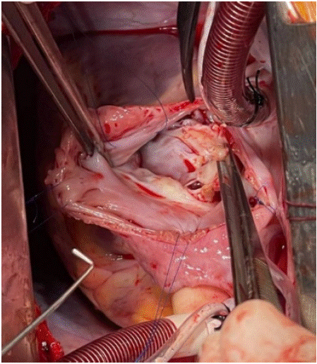
After a suspicion of infective endocarditis of the right side of the heart, the patient was treated with antibiotic triple therapy for a period of two weeks. Escherichia coli was isolated on haemocultures and the patient became afebrile after 5 days on antibiotics. After improvement in the renal parameters and haemoglobin levels he underwent cardiac surgery (Figure 1).

Figure 1: Exposure of the septal defect and the vegetation on the tricuspid valve septal leaflet.
The heart was approached in standard median sternotomy and cardio-pulmonary bypass was established by aortic and bi-caval cannulation. The heart was arrested with anterograde cardioplegia and the right atrium was opened. The septal leaflet of the tricuspid valve was full of vegetations and was resected.
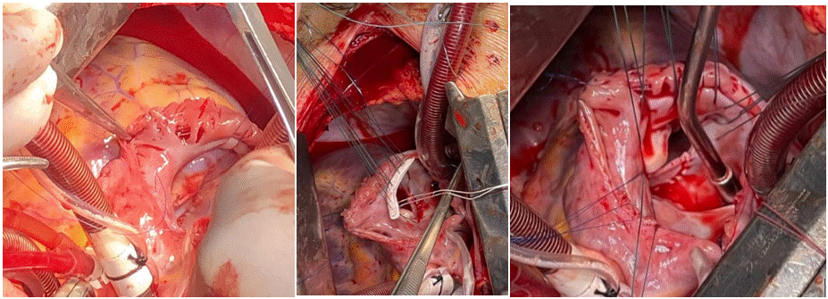
The ventricular defect was closed with autologous pericardial patch. The pericardium was used also to reconstruct the septal leaflet of the valve which was attached also to the native posterior leaflet realising bicuspidazion of the valve.
Artificial gore-tex neocordae tendinae and a tricuspid annular ring (plicated gore-tex tube 6 mm) were implanted to achieve competence of the valve on the hydraulic test. The right atrium was closed, the aorta declamped dhe the patient was weaned from the heart-lung machine. After TEE control the sternum was closed and he was transferred in the intensive care unit (Figure 2).

Figure 2: Repair of the tricuspid valve with pericardial patch and gore-tex annulus.
Postoperative recovery was uneventful and the patient left the hospital in good conditions ten days after surgery. He received antibiotics for another three weeks and on control laboratory data were normal except a slightly elevated serum creatinine. The tricuspid valve had minimal regurgitation and no residual septal defect was detected on echocardiography.
Discussion
Infective endocarditis (IE) has an annual incidence of 3–10/100,000 of the population with a mortality of up to 30% at 30 days [1,2]. In the era of modern medicine, healthcare-associated IE is gaining more and more terrain and accounts for 25–30% of contemporary reports as a result of a greater use of intravenous lines and intracardiac devices.
Right-sided IE is a minority in this group of critically ill patients and accounts for up to 10% of all patients with IE, but its frequency may be increasing in some countries due to more present risk factors like: patients with congenital heart disease, indwelling catheters, immunocompromised patients, etc [3,4]. Tricuspid and pulmonary valves may be involved in the setting of right sided infective endocarditis.
Congenital heart disease is the most common predisposing factor for paediatric infective endocarditis. Classically, patients with unrepaired ventricular septal defects (VSDs) are at greater risk of IE and closure may be a reasonable option in selected cases. Nowadays, the American Heart Association (AHA) considers VSDs to be relatively low risk and therefore does not recommend even antibiotic prophylaxis against IE.
A Danish population-based cohort study reported a high hazard ratio (HR) of endocarditis in patients with VSDs, both unrepaired and surgically closed [5]. But we can observe in the literature reports of series of children with small, restrictive, peri-membranous VSDs who developed tricuspid valve (TV) IE. They experience delayed diagnosis and secondary complications, including septic pulmonary emboli. These cases draw attention to the potential benefits of VSD closure in this group of children [6].
Regarding pathophysiological mechanisms, infective endocarditis of the trisucpid valve is directly correlated with turbulent flow through the defect. In a predisposing situation during life, a transient bacteraemia may lead to infection of the endocardium and formation of vegetations in the highest turbulence flow zone. In rare circumstances, when a small peri-membranous VSD is neglected during childhood it can be a starting point of triscuspid valve endocarditis in adults. A trigger like recurrent urinary tract infections with frequent bacteriemia in the terrain of this unresolved congenital heart defect can lead to infective endocarditis.
A conservative approach is recommended for the majority of patients with IE affecting the tricuspid or pulmonary valve. Recurrent pulmonary emboli are not an absolute indication for surgery. Surgery is indicated if fever persists despite 3 weeks of appropriate antibiotic treatment. Surgical options include debridement of the infected area, vegetation excision with either valve preservation or valve repair. If the repair is not possible, excision of the tricuspid valve with prosthetic valve replacement, is an enforced decision. Tricuspid valvectomy without use of a prosthesis has been advocated in extreme cases but may be associated with severe postoperative right-sided heart failure, particularly in patients with pulmonary hypertension as a result of multiple pulmonary emboli [7].
We think our case is very rare because the patient was neglected during childhood due to poor social and economic conditions of the family and presented to the hospital with acute renal failure in the terrain of right sided infective endocarditis.
A dedicated team available at a tertiary centre should manage patients with IE. This should comprise cardiologists, infectious diseases specialists and also cardiac surgeons. Since 30% of the patients will experience symptomatic neurological events, a neurologist will be of great help. This IE team approach enables early referral to surgery, appropriate antibiotic medication regimens, access to advanced imaging, close monitoring for complications and followup once treatment is complete. In this setting, 1-year mortality can be expected to be approximately halved [8].
Conclusion
Even nowadays in the era of modern medicine, we should not forget an unfollowed congenital heart disease as a predisposing condition for infective endocarditis. A multi-disciplinary team is effective in treating this complex pathology and achieve good results. We had a successful treatment of this patient especially in terms of surgery where we realised a good tricuspid valve repair.
References
- Mostaghim AS, Lo HYA and Khardori N. A retrospective epidemiologic study to define risk factors, microbiology, and clinical outcomes of infective endocarditis in a large tertiary-care teaching hospital. SAGE Open Med. 2017; 5: 2050312117741772.
- Murdoch DR, Corey GR, Hoen B, et al. Clinical presentation, etiology, and outcome of infective endocarditis in the 21st century: the International Collaboration on Endocarditis-Prospective Cohort Study. Arch Intern Med. 2009; 169: 463–473.
- Lassen H, Nielsen SL, Gill SUA, Johansen IS. The epidemiology of infective endocarditis with focus on non-device related right-sided infective endocarditis: a retrospective register-based study in the region of southern Denmark. Int J Infect Dis. 2020; 95: 224–230.
- Sridhar AR, Lavu M, Yarlagadda V, Reddy M, Gunda S, Afzal R, et al. Cardiac implantable electronic device-related infection and extraction trends in the U.S. Pacing Clin Electrophysiol. 2017; 40: 286–293.
- Eckerstrom F, et al. Lifetime burden of morbidity in patients with isolated congenital ventricular septal defect. J Am Heart Assoc. 2023; 12: e027477.
- Butensky AM, Channing A, Handel AS, et al. Tricuspid Valve Endocarditis in Four Patients with Unrepaired Restrictive Perimembranous Ventricular Septal Defects. Pediatr Cardiol. 2022; 43: 1929–1933.
- Musci M, Siniawski H, Pasic M, Grauhan O, Weng Y, Meyer R, et al. Surgical treatment of right-sided active infective endocarditis with or without involvement of the left heart: 20-year single center experience. Eur J Cardiothorac Surg. 2007; 32: 118 –125.
- Victoria Delgado, Nina Ajmone Marsan et al. 2023 ESC Guidelines for the management of endocarditis: Developed by the task force on the management of endocarditis of the European Society of Cardiology (ESC) Endorsed by the European Association for Cardio-Thoracic Surgery (EACTS) and the European Association of Nuclear Medicine (EANM), European Heart Journal. 2023; 44: 3948–4042.