
Case Report
Austin J Clin Case Rep. 2025; 12(1): 1348.
Major Complications of Implants Retaining Removable Prostheses - Case Reports and Proposal of a Novel Attachment Concept
Schulz A¹, Roth D², Orujov K¹, Karl M³*, Grobecker-Karl T¹
¹Department of Prosthodontics, Saarland University, Homburg/Saar, Germany
²Hader Solutions & Distribution, Swords/Co. Dublin, Ireland
³Department of Prosthodontics, Philipps University, Marburg, Germany
*Corresponding author: Matthias Karl, Department of Prosthodontics, Philipps University, Georg-Voigt Strasse 3, 35039 Marburg, Germany. Tel: +49 6421 5863277; Fax: +49 6421 5862904. Email: karl_matthias@web.de
Received: January 07, 2025; Accepted: January 28, 2025; Published: February 03, 2025
Abstract
Removable restorations on implants and natural abutments constitute a viable treatment option with telescopic crowns often used as attachments. In two cases, catastrophic failures occurred, both being attributable to mechanical overloading. In the first patient, an implant fracture occurred under a removable cantilever prosthesis while in the second patient an implant was lost which had been retrofitted with a locator attachment to support a preexisting prosthesis. A novel attachment system, incorporating a flexible element in the male component is introduced which compensates for fabrication inaccuracies and non-parallelism of supporting implants, which might have acted as stressbreaker thereby preventing implant overload.
Keywords: Implant fracture; Loss of osseointegration; Locator
Introduction
Treatment with implant-supported overdentures has been repeatedly shown to increase patient comfort and quality of life [1,2]. Major complications reported in such restorations include periimplantitis [3] and technical problems with incidences of 11.4% for abutment fractures and 3.5% for implant fractures [4].
Ball anchors and locator abutments have been widely employed as attachment systems [5,6] with loss of retention [7,8], wear of female retentive components [9,10] and fractures of the removable prostheses [7,11] constituting the most frequently occurring problems. According to a systematic literature review, these mechanical failures have similar prevalence as compared to biologic and esthetic failures [12].
Telescopic crowns [1] have been described as common denominator for combining implants and natural abutments for supporting removable prostheses. In contrast to prefabricated attachments, telescopic crowns can be placed on both, implants and teeth. It is known that high moment loads [13,14] occur with rigid telescopic crowns supporting prostheses moving under masticatory loads, which may lead to implant fractures [15] due to increased stress in the implant body which is even worse when peri-implant bone resorption already has occurred [16]. A current report showed a tendency towards a greater incidence of complications for implants restored with removable restorations [17], which is in line with a recently published retrospective analysis of telescopic crown supported prostheses describing that 2.3% of implants had failed [1].
Establishing a common path of insertion [18-20] of attachments is considered as being relevant for achieving long term success [21]. This may either be realized by individually fabricating telescopic crowns [1,13] or by exploiting the geometric form [19] of singlestanding, prefabricated attachments [22] as well as by using various retentive inserts made from different types of plastic materials [23]. Several authors have reported that lacking a common path of insertion resulted in compromised retention and accelerated wear of attachment systems [8,20,24,25]. In this context, salvaging an existing removable prosthesis by retrofitting an implant and a single standing attachment constitutes a special situation, which for instance occurs when a natural abutment has fractured requiring extraction [26]. In such cases, a common path of insertion of pre-existing telescopic crowns and ball anchor or locator cannot be established.
In addition, inevitable positional errors during fabrication of superstructures prevent a passive fit being achieved [27]. While bone adaptation minimizing misfit between implants and restoration has been proven for fixed restorations [28], a literature review described component fractures and loss of implants as potential consequences of misfitting superstructures [27]. In the same context, a retrospective clinical study revealed marginal bone loss in one third of the implant length or more in 19% of implants supporting overdentures [29], which may also be attributed to the static and dynamic loading conditions present in these cases.
This article reports on a patient restored with a removable restoration on implants and teeth employing telescopic crowns for retention who fractured an implant following previous technical complications. In a second patient, a locator abutment was used for salvaging an existing telescopic crown supported, removable prosthesis, which led to implant loss after three years. Using qualitative photoelastic analysis, the potential of a novel attachment system [30,31] incorporating a flexible element into the male component for acting as a stress-breaker preventing implant overload was evaluated.
Case Presentation – Implant Fracture
A male patient initially presented in September 2020 at the age of 76 with implant-supported fixed restorations in the mandible (Figure 1a). Fixed restorations on implants and teeth were present in the maxilla and the patient required extractions of posterior teeth due to abscesses. Following healing, the patient was treatment planned for a combined tooth-implant supported removable restoration in January 2021 following implant placement in the position of tooth 15 (Figures 1b, 1c). In November 2021 a prosthesis employing telescopic crowns supported by both natural abutments and implants was delivered (Figure 2).
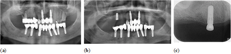
    Figure 1: Panoramic radiograph showing implant supported restorations in the mandible while both, teeth and implants were present in the maxilla supporting
fixed restorations. Abscesses had developed involving posterior teeth on both sides (a). Radiograph following implant placement in the region of the maxillary
right second premolar (b). Following osseointegration, the implant was uncovered and a healing cap was installed (c).
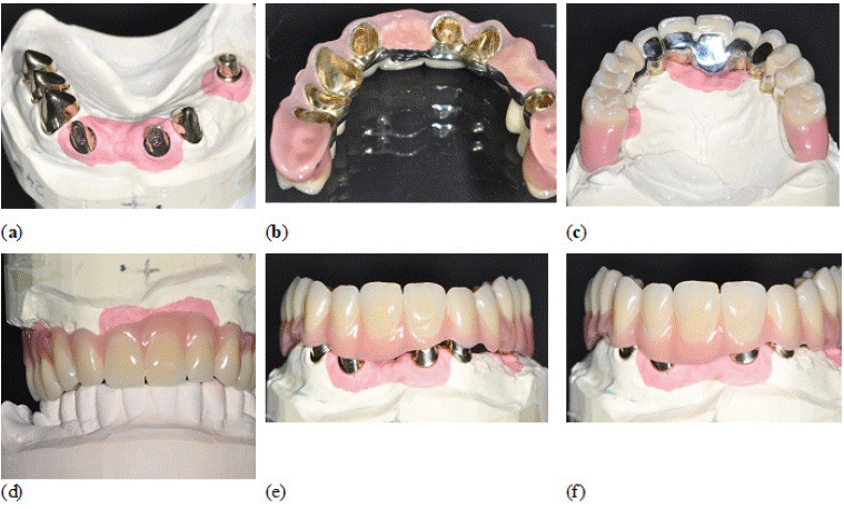
    Figure 2: A total of four telescopic crowns on natural abutments and three additional telescopic crowns formed by implant abutments (a) were used to support
a horseshoe-shaped removable restoration (b) with gracile dimensions in the anterior region (c). Given the patient’s wish not to alter the pre-existing mandibular
restorations, an ideal plane of occlusion could not be established (d). The cylindrical telescopic crowns were fabricated by casting high-noble gold alloy and
milled such way that a smooth insertion process could be ensured (e,f).
In September 2022 the patient presented with a fracture of the prosthesis framework (Figure 3) requiring major repair followed by a chipping fracture of an anterior facing which occurred in November 2022 (Figure 4). Ultimately, in March 2023 the implant in position 15 fractured (Figures 5, 6) with the patient reporting to our clinic as he had been unable to properly position the prosthesis for several days. As the patient was still satisfied with the function and retention of the removable restoration, it was decided not to remove the implant but to smooth it and to extend the base of the prosthesis in this area.
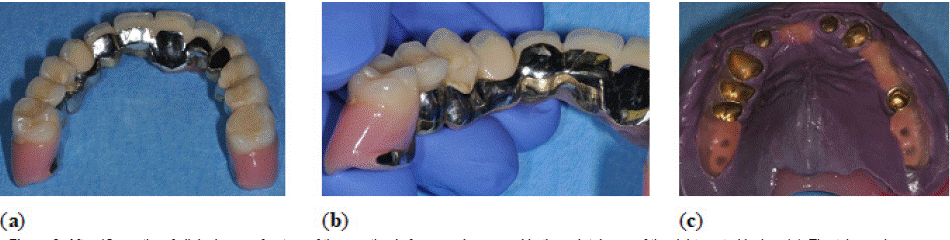
    Figure 3: After 10 months of clinical use, a fracture of the prosthesis framework occurred in the palatal area of the right central incisor (a). The telescopic crowns
had been bonded to the non-noble alloy framework (b) and after repositioning of the removable prosthesis, an impression was made using polyether material (c).
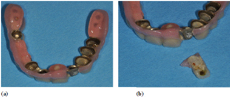
    Figure 4: The facing of the left lateral incisor fabricated from composite resin
(a) chipped off the framework requiring renewal (b). The plaque formed on
the internal surface of the facing indicates that a gap had been existing for a
longer period of time.
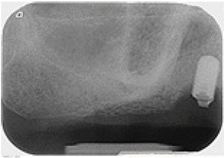
    Figure 5: Periapical radiograph of implant in position 15 following fracture of
the coronal part. A saucer-shaped bone defect is clearly visible surrounding
the implant.
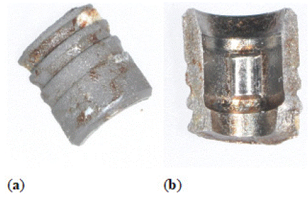
    Figure 6: Only one part of the fractured implant (a) was still present when
the patient reported to our clinic consisting of approximately 50% of the
circumference of the implant abutment connection (b).
Case Presentation – Implant Loss
A 66 year old male patient initially presented in 2011 having been restored elsewhere with conventional tooth-supported fixed restorations and showing a mix of periodontal and endodontic problems (Figure 7). Following implant placement in positions 13 and 25 in June 2012, a removable, horseshoe-shaped maxillary prosthesis on telescopic crowns was delivered in February 2013 (Figure 8). As part of the maintenance program, the patient was seen on a regular basis only requiring minor interventions such as cementing loose primary telescopic crowns.
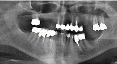
    Figure 7: Initial patient presentation with partial edentulism and preexisting
fixed restorations in both jaws
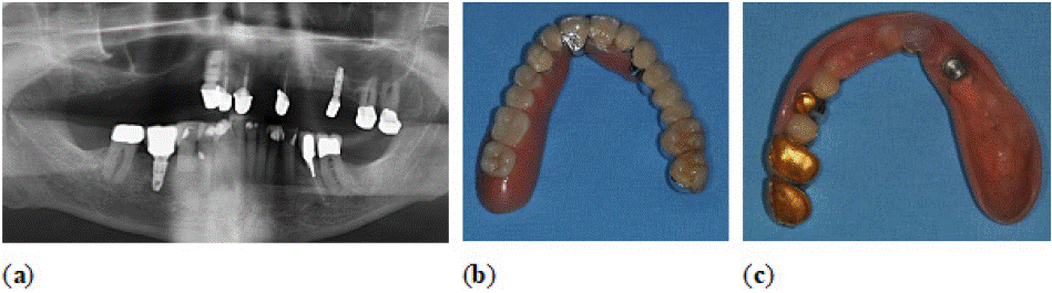
    Figure 8: Panoramic radiograph (a) after maxillary treatment with two implants in the region of tooth 13 and tooth 25. The patient was restored with a removable
horseshoe-shaped prosthesis supported by cylindrical telescopic crowns (b) which was extended whenever abutments were removed and ultimately was
retrofitted with a housing for a locator abutment (c).
In October 2020, tooth 12 had to be extracted due to a horizontal fracture (Figure 9a) and the primary telescopic crown on implant 13 had been lost by the patient. In order to salvage the existing prosthesis, the abutment on implant 13 was removed (Figure 9b) and ultimately replaced by a locator abutment in September 2021. The female attachment part was then retrofitted into the prosthesis (Figure 8c). In September 2023, tooth 11 fractured and had to be extracted (Figure 10a) with the preoperative radiograph revealing an osteolysis around implant 13 correlating with its clinical mobility (Figure 10b). The implant was ultimately removed (Figure 11) and the prosthesis modified such way that the palate was completely covered for additional support.
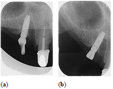
    Figure 9: Approximately seven years after initial rehabilitation, tooth 12
had to be removed due to a horizontal fracture and the primary telescopic
crown on implant 13 had been lost by the patient (a). The custom made
abutment on implant 13 was replaced by a healing abutment (b) which was
subsequently replaced by a locator followed by retrofitting the prosthesis.
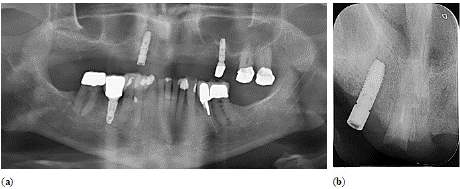
    Figure 10: Two years after prosthesis conversion, tooth 11 fractured and had
to be extracted (a). The preoperatively taken periapical radiograph revealed
an osteolysis surrounding implant 13 which was also clinically mobile (b).
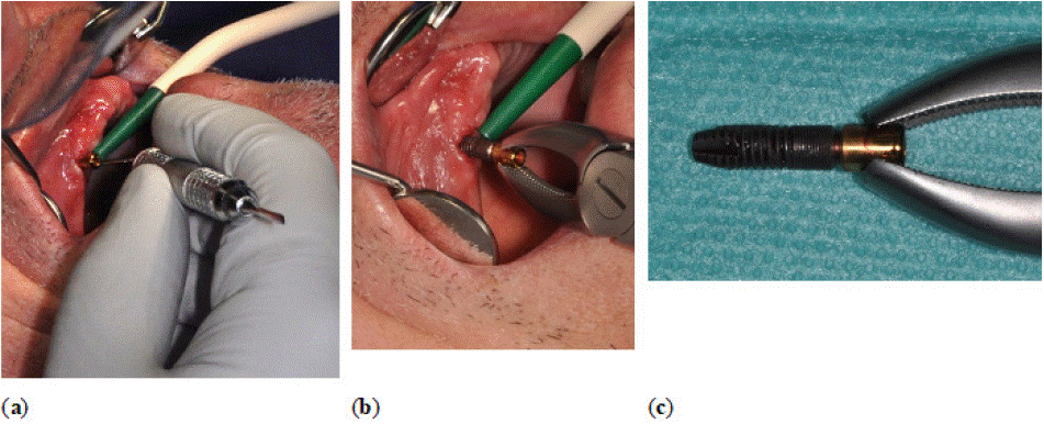
    Figure 11: Using a periotome, the soft tissue was carefully separated from the implant (a) which was then removed using forceps (b). No remnants of bone were
visible on the implant surface (c).
Photoelastic Study
Two bone-level implants (Straumann BLT RC 4.1x10 mm, Straumann AG, Basel, Switzerland) were mounted in a sheet of photoelastic resin (Figure 12) made from polycarbonate (Tiedemann Instruments, Garmisch-Partenkirchen, Germany) serving as a simulated patient situation [32-34]. Following open-tray impression making with polyether material (Impregum, 3M, Seefeld, Germany) and transfer components, a working cast (Fujirock, GC Germany, Bad Homburg, Germany) resembling the simulated patient was obtained, which was then used for manufacturing a bar-shaped removable prosthesis using acrylic resin (ProBase Cold, Ivoclar Vivadent, Schaan, Liechtenstein). An orthodontic expansion screw was incorporated into the restoration allowing to increase its overall length simulating different levels of prosthesis misfit. The restoration was fabricated to either fit a one-piece, stiff ball anchor (Clix Abutment, Hader Solutions & Distribution, Applewood, Ireland) or an identically sized flexible attachment (Reflex, Hader Solutions & Distribution) both of which could be mounted on the implants (Figure 13) [30,31].
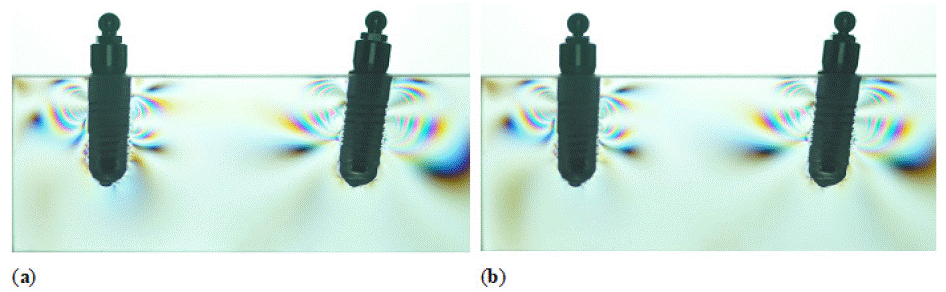
    Figure 12: Model situation made from photoelastic resin with two bone level implants onto which a stiff, one-piece ball anchor (a) or a flexible attachment
(b) could be mounted.
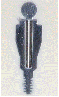
    Figure 13: Cross-sectional view of a prototype flexible attachment system
(Reflex, Hader Solutions & Distribution) with a ball mounted on a centrally
positioned beam allowing for lateral movement while maintaining vertical
dimensions.
The bar-shaped prosthesis was attached to the simulated patient situation and placed in a polariscope (Tiedemann Instruments) for photoelastic analysis [32-34]. Standardized photographs were made allowing for qualitative analysis of stress patterns indicating the contours of principal stress differences. This method has been described as simple technique for rough estimations on stress intensity and stress concentrations based on colour, number and closeness of emerging fringe patterns [32-34].
The use of the one-piece Clix abutments led to the apparition of additional visible deformations of the photoelastic resin indicating additional strain in the area between both implants and bellow their apices (Figure 14b) when prosthesis misfit was simulated by opening the orthodontic screw. This was due to the direct transmission of the strain to the implant through the stiff abutments and obviously misfit was present in the restoration even without expanding the orthodontic screw resulting from fabrication inaccuracies (Figure 14a).
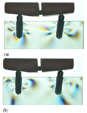
    Figure 14: Seating of the bar on the ball anchors already evoked stress in
the bone surrounding both implants (a) which was due to inherent misfit. This
situation worsened with opening of the orthodontic screw incorporated in the
bar thereby increasing the amount of misfit (b).
In the same situations (Figure 15a), the Reflex abutments compensated for positional mismatch between implants and prosthesis. This led to a preservation of the area around the implants and was confirmed by only a slight increase of the deformations following expansion of the orthodontic screw (Figure 15b).
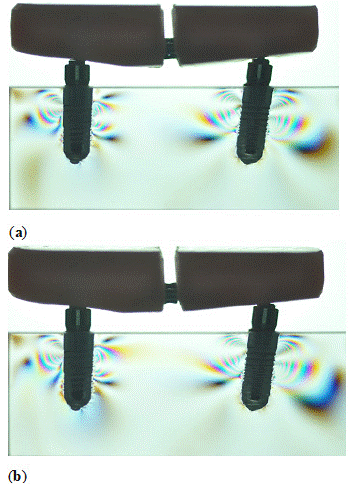
    Figure 15: When the bar was seated on the flexible attachment system,
only minor stress was seen surrounding the supporting implants (a) and this
hardly changed by increasing the misfit of the bar.
Discussion/Conclusions
Two similar cases of major complications occurring in patients restored with telescopic crown supported removable restorations on natural and implant abutments have been described. In both cases, implants were used to supplement remaining teeth for additional support and retention of the prostheses what has been recommended as a reasonable treatment approach [1]. Based on a broad database, a greater incidence of complications for implants restored with removable restorations [17] has been shown, what is not surprising given their unfavorable biomechanical situation. Especially technical complications encompassing both, implant components [4] and removable prostheses [7,11] seem to be quite frequent in implant supported removable restorations [12] and hence the cases presented here fit into that scheme. In both cases, the implants were the distal most support of cantilevers resulting in considerable moment loading [13,15]. The massive peri-implant bone loss observed in the patient with the fractured implant may also be attributed to an overload situation and has similarly been observed in a well documented clinical study [29].
Apart from the loading situation of the prostheses under masticatory function, it can be argued that misfit was also present in both patients’ removable restorations leading to localized stress. Misfit has been shown to be mostly due to transfer inaccuracies between the patient situation and the laboratory situation as well as dimensional changes of restorative materials [27,28]. In the patient with the fractured implant, these stresses may have contributed to the fracture of the reinforcing framework and the subsequent chipping of the facing. In the second patient who lost an implant, the static loading situation resulting from misfit may have caused overload of alveolar bone [27].
A specific problem for the patient with the retrofitted locator attachment can be seen in a lack of achieving a common path of insertion of the pre-existing telescopic crowns and the newly added locator [18-20]. While achieving a common path of insertion in telescopic crowns is achieved by manual milling in the dental laboratory making these very cost-intense attachments, prefabricated attachments either exploit their geometric form [19] or use various retentive inserts [23] for achieving this goal.
The two clinical cases presented sparked the idea of establishing a novel attachment system [30], which allows to compensate for misalignments of implants as well as for inevitable transfer inaccuracies occurring during superstructure fabrication. These two factors have to be seen as the main reasons for the major problems described for implant-supported overdentures on prefabricated single-standing attachments i.e. prosthesis fracture and component wear [7-11]. Ideally, such an attachment system would also act as stress breaker preventing implant overload even in cantilever situations as seen in the clinical cases. As evidenced in the photoelastic study, the central beam inside the abutment supporting the retentive ball allows for sufficient compensatory movement in order to minimize peri-implant bone loading, which is in contrast to current attachment systems employing different female inserts made from plastic materials.
References
- Zierden K, Wöstmann B, Rehmann P. Which Patient-Related Factors Influence the Outcome of Telescopic-Retained Removable Implant-Supported Dental Prostheses in Edentulous Patients? Int J Prosthodont. 2022; 35: 690-696.
- Prasad S, Faverani LP, Santiago Junior JF, Sukotjo C, Yuan JC. Attachment systems for mandibular im-plant-supported overdentures: A systematic review and meta-analysis of randomized controlled trials. J Prosthet Dent. 2024; 132: 354-368.
- Rammelsberg P, Bernhart G, Lorenzo Bermejo J, Schmitter M, Schwarz S. Prognosis of implants and abutment teeth under combined tooth-implantsupported and solely implant-supported double-crown-retained removable dental prostheses. Clin Oral Implants Res. 2014; 25: 813-818.
- Yi Y, Heo SJ, Koak JY, Kim SK. Mechanical complications of implantsupported restorations with internal conical connection implants: A 14-year retrospective study. J Prosthet Dent. 2023; 129: 732-740.
- Kleis WK, Kämmerer PW, Hartmann S, Al-Nawas B, Wagner W. A comparison of three different attachment sys-tems for mandibular twoimplant overdentures: one-year report. Clin Implant Dent Relat Res. 2010; 12: 209-218.
- Türk PE, Geckili O, Türk Y, Günay V, Bilgin T. In vitro comparison of the retentive properties of ball and locator attachments for implant overdentures. Int J Oral Maxillofac Implants. 2014; 29: 1106-1113.
- Parzham V, Judge RB, Bailey D. A 5-Year Retrospective Assay of Implant Treatments in Private Practice: The Restor-ative Complications of Long- Span Implant-Supported Fixed and Removable Dental Prostheses. Int J Prosthodont. 2020; 33: 493-502.
- Rabbani S, Juszczyk AS, Clark RK, Radford DR. Investigation of retentive force reduction and wear of the locator attachment system with different implant angulations. Int J Oral Maxillofac Implants. 2015; 30: 556-563.
- ELsyad MA, Abdraboh AE, Denewar MM, Mohamed SS. Prosthetic complications and maintenance of different attachments used to stabilize mandibular 2-implant overdentures in patients with atrophied ridges. A 5-year random-ized controlled clinical trial. Clin Implant Dent Relat Res. 2022; 24: 497-509.
- Friedrichsen M, Dirksen D, Runte C. In vitro measurement of the retention force of two stud attachment systems during cyclic load. J Prosthodont. 2024; 33: 164-170.
- de Paula MS, Cardoso JB, de Menezes EEG, Nogueira TE, McKenna G, Leles CR. A prospective cohort on the incidence of fractures in single-implant mandibular overdentures. J Dent. 2020; 103: 103521.
- Verma A, Singh SV, Arya D, Shivakumar S, Chand P. Mechanical failures of dental implants and supported prostheses: A systematic review. J Oral Biol Craniofac Res. 2023; 13: 306-314.
- Heckmann SM, Winter W, Meyer M, Weber HP, Wichmann MG. Overdenture attachment selection and the loading of implant and denture-bearing area. Part 2: A methodical study using five types of attachment. Clin Oral Implants Res. 2001; 12: 640-647.
- Grobecker-Karl T, Kafitz L, Karl M. Effect of Implant Position and Attachment Type on the Biomechanical Behavior of Mandibular Single Implant Prostheses. Eur J Prosthodont Restor Dent. 2020; 28: 152-160.
- Karl M, Krafft T, Kelly JR. Fracture of a narrow-diameter Roxolid implant: clinical and fractographic considerations. Int J Oral Maxillofac Implants. 2014; 29: 1193-1196.
- Bing L, Mito T, Yoda N, Sato E, Shigemitsu R, Han JM, Sasaki K. Effect of peri-implant bone resorption on mechanical stress in the implant body: In vivo measured load-based finite element analysis. J Oral Rehabil. 2020; 47: 1566-1573.
- Rammelsberg P, Lorenzo-Bermejo J, Kappel S. Effect of prosthetic restoration on implant survival and success. Clin Oral Implants Res. 2017; 28: 1296- 1302.
- Choi JW, Bae JH, Jeong CM, Huh JB. Retention and wear behaviors of two implant overdenture stud-type at-tachments at different implant angulations. J Prosthet Dent. 2017; 117: 628-635.
- Srinivasan M, Kalberer N, Maniewicz S, Müller F. Implant-Retained Overdentures Using an Attachment with True-Alignment Correction: A Case Series. Int J Prosthodont. 2019; 32: 482-496.
- Ortegón SM, Thompson GA, Agar JR, Taylor TD, Perdikis D. Retention forces of spherical attachments as a func-tion of implant and matrix angulation in mandibular overdentures: an in vitro study. J Prosthet Dent. 2009; 101: 231- 238.
- Al-Ghafli SA, Michalakis KX, Hirayama H, Kang K. The in vitro effect of different implant angulations and cyclic dislodgement on the retentive properties of an overdenture attachment system. J Prosthet Dent. 2009; 102: 140-147.
- Stephens GJ, di Vitale N, O’Sullivan E, McDonald A. The influence of interimplant divergence on the retention characteristics of locator attachments, a laboratory study. J Prosthodont. 2014; 23: 467-475.
- ELsyad MA, Emera RM, Ashmawy TM. Effect of Distal Implant Inclination on Dislodging Forces of Different Loca-tor Attachments Used for Mandibular Overdentures: An In Vitro Study. J Prosthodont. 2019; 28: e666-e674.
- Passia N, Ghazal M, Kern M. Long-term retention behaviour of resin matrix attachment systems for overdentures. J Mech Behav Biomed Mater. 2016; 57: 88-94.
- https://pubmed.ncbi.nlm.nih.gov/21426402/
- Rauch A, Hahnel S, Köthe S, Schierz O. Improving Oral Health-Related Quality of Life by Converting Fractured Abutment Teeth in Double Crown- Retained Removable Prostheses into Root-Anchored Ball Attachments. Int J Prosthodont. 2019; 32: 389-392.
- Katsoulis J, Takeichi T, Sol Gaviria A, Peter L, Katsoulis K. Misfit of implant prostheses and its impact on clinical outcomes. Definition, assessment and a systematic review of the literature. Eur J Oral Implantol. 2017; 10 Suppl 1: 121-138.
- Karl M, Taylor TD. Bone Adaptation Induced by Non-Passively Fitting Implant Superstructures: A Randomized Clinical Trial. Int J Oral Maxillofac Implants. 2016; 31: 369-375.
- Marinis A, Afshari FS, Yuan JC, Lee DJ, Syros G, Knoernschild KL, et al. Retrospective Analysis of Implant Overdenture Treatment in the Advanced Prosthodontic Clinic at the University of Illinois at Chicago. J Oral Implantol. 2016; 42: 46-53.
- Grobecker-Karl T, Kafitz L, Karl M. Rationale for a novel attachment system for implant-supported overdentures. Int J Prosthodont. 2022; 35: 74–81.
- Wendler F, Diehl L, Shayanfard P, Karl M. Implant-Supported Overdentures: Current Status and Preclinical Testing of a Novel Attachment System. J Clin Med. 2023; 12: 1012.
- Karl M, Dickinson A, Holst S, Holst A. Biomechanical methods applied in dentistry: a comparative overview of photoelastic examinations, strain gauge measurements, finite element analysis and three-dimensional deformation analysis. Eur J Prosthodont Restor Dent. 2009; 17: 50-57.
- Verma V, Hazari P, Verma P. Biomechanical efficiency of different implantabutment connection: a systematic re-view of studies using photoelastic stress analysis. Evid Based Dent. 2023; 24: 92.
- Tonin BSH, Peixoto RF, Fu J, Freitas BN, Mattos MDGC, Macedo AP. Evaluation of misfit and stress distribution in implant-retained prosthesis obtained by different methods. Braz Dent J. 2021; 32: 67-76.