
Case Report
Austin J Clin Case Rep. 2025; 12(1): 1347.
Intrathoracic Migration of a Broken Port Needle: An Unusual Complication and Management
Jain R and Donuru A*
Department of Radiology, Hospital of the University of Pennsylvania, Philadelphia, PA, 19104, USA
*Corresponding author: Achala Donuru, MBBS, FRCR, Associate Professor of Radiology, Hospitals of University of Pennsylvania, 3400 Spruce Street, Philadelphia, PA, 19104, USA Tel: 215.662.3036; Fax: 215.614.0033; Email: achala. donuru@pennmedicine.upenn.edu
Received: January 06, 2025; Accepted: January 28, 2025; Published: January 31, 2025
Abstract
Central venous port devices are commonly used in oncology patients requiring long-term intravenous therapy. While generally safe, complications can occur. We present a case of a fractured port needle that rapidly migrated intrathoracically, resulting in a large pneumothorax. The needle was successfully retrieved via a combined thoracoscopic and bronchoscopic approach. This case highlights the importance of prompt recognition and management of rare complications associated with port placement.
Keywords: Central venous port; Needle fracture; Migration; Pneumothorax; Thoracoscopy; Bronchoscopy: Case report
Case Presentation
A 42-year-old male with a history of recurrent metastatic testicular cancer presented for port placement in preparation for chemotherapy. He had previously undergone orchiectomy and retroperitoneal lymph node dissection twenty years ago and had recently developed a new osseous lesion in the sternum confirmed to be metastatic on biopsy.
After obtaining informed consent, the port placement procedure was initiated. During the procedure, a 25-gauge needle fractured within the subcutaneous tissue, with an estimated length of 3.5 cm. Initial attempts to retrieve the needle fragment were unsuccessful. Shortly thereafter, while ambulating to the restroom, the patient experienced a sudden onset of intense, sharp chest pain and shortness of breath. Physical examination revealed decreased breath sounds in the left upper lung field.
An immediate chest radiograph demonstrated a new small leftsided pneumothorax and a linear hyperdense foreign body in the left lower zone, located in the para-mediastinal region (Figure 1). The findings were promptly discussed with the thoracic surgery team. A repeat radiograph demonstrated an enlarging left pneumothorax (Figure 2). An emergent computed tomography (CT) scan of the chest was performed for further characterization and localization of the foreign body. The CT revealed a large left pneumothorax with a contralateral mediastinal shift. A 3.5 cm radiopaque linear foreign body, consistent with the broken needle, was identified along the left pericardial sac, extending intrathoracically (Figure 3, 4).
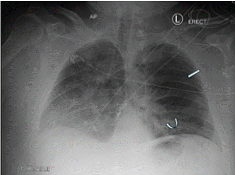
    Figure 1: Chest radiograph demonstrates a small left-sided pneumothorax
(straight arrow) and a linear hyperdense foreign body in the left lower zone
(curved arrow).
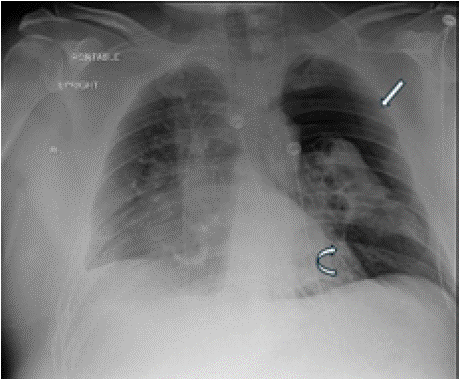
    Figure 2: Repeat chest radiograph demonstrates a moderate to large leftsided
pneumothorax (straight arrow) and a linear hyperdense foreign body
in the left lower zone (curved arrow).
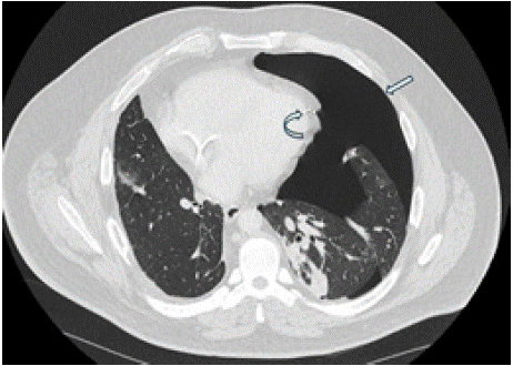
    Figure 3: CT scan axial image showing large left pneumothorax (straight
arrow) with contralateral mediastinal shift. The radiopaque linear foreign
body is noted in the left pericardial sac (curved arrow)
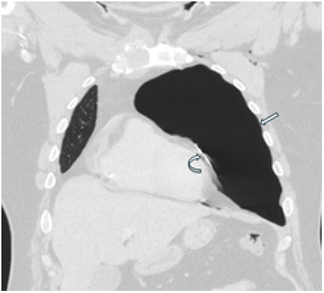
    Figure 4: CT scan coronal image demonstrates a large left pneumothorax
(straight arrow) and a radiopaque linear foreign body along the left
pericardial sac (curved arrow).
Urgent combined bronchoscopy and thoracoscopy were performed. Bronchoscopy was utilized to visualize the tracheobronchial tree and rule out airway injury. The patient was then positioned in the left lateral decubitus position, and thoracoscopic ports were placed. The foreign body was successfully retrieved using a laparoscopic needle driver. The retrieved fragment measured 3.5 cm and was confirmed to be the broken needle.
Post-procedural imaging demonstrated complete resolution of the left pneumothorax (Figure 5). The patient's symptoms improved significantly, and no further complications were noted.
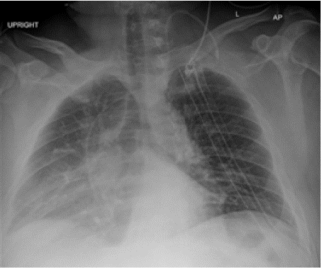
    Figure 5: Post-procedural imaging demonstrated complete resolution of the
left pneumothorax. Two left chest drains are noted.
Discussion
Initially introduced in 1982, central venous port systems have become indispensable in modern oncologic care. The reported overall complication rate for these devices ranges from 7.2% to 12.5%, with port-system fractures occurring in 0.4% to 1.8% of cases [1-3]. Complications can be categorized as early (within 30 days) or delayed (after 30 days) and as minor or major, with the latter requiring intervention within 24 hours [1].
Factors contributing to port catheter fracture include the type of catheter, site of placement, and duration of use [4]. Catheter rupture typically occurs as a delayed complication, often in the space between the first rib and clavicle due to mechanical friction. Migration of fractured fragments can be influenced by vigorous upper arm movement, neck flexion, and changes in intrathoracic pressure associated with coughing, straining, or vomiting [2]. The incidence of pneumothorax following port catheter placement varies between 1.5% and 6% [1,5]. While intraoperative fluoroscopy is useful, its limited field of view may not reliably exclude pneumothorax, making a post-procedural chest radiograph essential [1].
In this case, the needle fractured during the initial stages of the procedure. The subsequent rapid migration likely resulted from a combination of patient movement, vascular flow, and straining, which propelled the sharp needle fragment into the pericardial space and eventually intrathoracically, causing lung injury and a progressive pneumothorax.
Prompt detection and accurate characterization of such complications are crucial for appropriate management. A routine post-procedural chest radiograph is vital, as it not only detected the pneumothorax in this case but also identified the migrated foreign body. CT imaging provides further precise localization and aids in evaluating associated complications, facilitating treatment planning.
Early removal of the fractured fragment is recommended to prevent further complications. Retrieval options include surgical intervention, percutaneous endovascular techniques, and, in rare cases, bronchoscopy. In this instance, a combined thoracoscopic and bronchoscopic approach successfully retrieved the needle fragment. Post-procedural imaging confirmed near-complete resolution of the pneumothorax, with significant symptomatic improvement.
Conclusion
Needle fracture and migration are rare but potentially serious complications of central venous port placement. This case highlights the importance of recognizing this complication, understanding the potential for rapid migration, and the need for prompt intervention. Imaging, particularly the initial chest radiograph, plays a crucial role in diagnosis, guiding management, and assessing treatment response. Clinicians should be aware of this potential complication and maintain a high index of suspicion, especially when patients present with acute chest pain or respiratory distress following port placement procedures. Prompt removal of migrated fragments is essential to prevent further morbidity.
References
- Machat S, Eisenhuber E, Pfarl G, Stübler J, Koelblinger C, Zacherl J. Complications of central venous port systems: a pictorial review. Insights Imaging. 2019; 10: 86.
- Kakkos A, Bresson L, Hudry D, Cousin S, Lervat C, Bogart E, et al. Complication-related removal of totally implantable venous access port systems: does the interval between placement and first use and the neutropenia-inducing potential of chemotherapy regimens influence their incidence? A four-year prospective study of 4045 patients. European Journal of Surgical Oncology (EJSO). 2017; 43: 689-695.
- Doley RP, Gupta D, Tigari P, Thapa P, Deka P, Kumar A. Catheter fracture and migration of a totally implantable venous access device: a case report. J Med Case Rep. 2014; 8: 112.
- Ko SY, Park SC, Hwang JK, Kim SD. Spontaneous fracture and migration of catheter of a totally implantable venous access port via internal jugular vein-a case report. Journal of Cardiothoracic Surgery. 2016; 11: 50.
- McGee DC, Gould MK. Preventing complications of central venous catheterization. New England journal of medicine. 2003; 348: 1123-1133.