
Case Report
Austin J Cerebrovasc Dis & Stroke. 2020; 7(1): 1085.
DWI Negative Large Artery Acute Ischemic Stroke: A Case Report with Review of Literature
Raut TP1*, Patil LN1, Munshi M2, Shrivastava M3, Sanghani G3, Shah P3
1Department of Neurosciences, Kokilaben Dhirubhai Ambani Hospital, Mumbai, India
2Department of Neuroradiology, Kokilaben Dhirubhai Ambani Hospital, Mumbai, India
3Department of Interventional Neuroradiology, Kokilaben Dhirubhai Ambani Hospital, Mumbai, India
*Corresponding author: Raut TP, Department of Neurosciences, Kokilaben Dhirubhai Ambani Hospital, Mumbai, India
Received: May 01, 2020; Accepted: October 09, 2020; Published: October 16, 2020
Abstract
MRI forms an important investigation tool in acute ischemic stroke. Diffusion weighted imaging is a very sensitive sequence to detect ischemia within few minutes. However in certain cases it can be negative especially in lacunar strokes, brainstem strokes and rarely in large artery strokes imaged very early into the onset of stroke. Other tools like MR Angiogram, Perfusion imaging may increase the sensitivity to detect stroke and initiate timely treatment which could prevent devastating consequences. Here authors describe a case of a 57 year old gentleman presenting with right sided weakness of 90 minutes duration with a moderate stroke severity. MRI Diffusion was negative. MR Angiogram didn’t reveal any obvious intracranial occlusion. However MR Perfusion revealed an ischemic penumbra in left anterior cerebral artery territory. Patient didn’t respond to iv thrombolysis and hence subjected to cerebral DSA which revealed thrombus and occlusion of left aca branch. Post mechanical thrombectomy with a stent retriever, he improved clinically and was functionally independent 2 weeks post discharge.
Keywords: Acute ischemic stroke; Diffusion weighted imaging; Magnetic resonance imaging; Perfusion Imaging
Abbreviations
MRI: Magnetic Resonance Imaging; CT: Computed Tomography; DWI: Diffusion Weighted Imaging; ADC: Apparent Diffusion Coefficient; FLAIR: Fluid Attenuated Inversion Recovery Sequence; PWI: Perfusion Weighted Imaging; DSA: Digital Subtraction Angiography; IV: Intravenous; AIS: Acute Ischemic Stroke; EVT: Endovascular Treatment
Introduction
Time is brain. Acute Ischemic Stroke (AIS) if untreated could be a debilitating and life threatening condition hence rapid diagnosis and treatment is necessary for optimizing the outcomes. Traditionally CT Brain has been used in emergency evaluation of patients with AIS to rule out intra-cranial bleed and selects patients for IV thrombolysis. Chalela et.al demonstrated Magnetic Resonance Imaging (MRI) to be more sensitive than CT in detecting acute ischemic stroke [1]. Recently there has been a significant increase in use of MRI in evaluation of patients with AIS [2]. At few centers MRI is used as primary screening tool to select patients eligible for thrombolysis or Endovascular Treatment (EVT). Particularly diffusion weighted magnetic resonance images compared to CT have better sensitivity, accuracy and interrater homogeneity [3]. Sensitivity of Diffusion Weighted Magnetic Resonance Images (DW-MRI) for detecting acute ischemic is stated to be 88-100% [4,5] and specificity 95-100% [4]. MRI-based thrombolysis is safer and potentially more efficacious than standard CT-based thrombolysis [6]. However, DW-MRI has its limitations and false negative DWI images in AIS have been described [7]. We report a case in which DWI-MRI was negative after 90 minutes of symptom onset and perfusion MRI images demonstrated early ischemia. Patient was aggressively treated and had a complete clinical recovery.
Case Presentation
A 57-year-old man, right-handed, businessman by occupation presented to the emergency with sudden onset weakness in right side of body of 90 minutes duration with absolute inability to move right lower limb. There was no history of backpain,radicular pain,fall,seizure or syncope. He had diabetes mellitus and hypertension and was on regular medications. On clinical examination BP was 120/80mmhg, Pulse 82/min, regular. Cranial nerve examination was normal, right upper limb power was MRC (Medical Research Council) grade 3/5 and right leg power MRC grade 0/5 with hypoesthesia on right side. Stroke severity as per NIHSS (National Institutes of Health Stroke Scale) was 7.
MRI Brain stroke protocol done showed negative DWI (Figure 1: A and B) and Fluid attenuated images (FLAIR) (Figure 2A). MRA Angiogram didn’t reveal any obvious extracranial or intracranial occlusion (Figure 2B). MR perfusion study done showed increased time to peak (Tmax) and Mean Transit Time (MTT) in para-sagittal left frontal lobe including paracentral lobule and peri-rolandic region suggestive of tissue at risk of ischemia (Figure 3 A and B). RAPID software showed significant diffusion and perfusion mismatch (Figure 3C). In view of significant deficit with ischemic penumbra despite normal diffusion, patient was considered eligible for IV Thrombolysis with alteplase and 0.9mg/kg tpa was administered. Patient failed to show improvement at the end of 1 hour. Emergent Digital subtraction angiography done showed left distal Anterior Cerebral Artery (ACA) thrombus, thrombotic occlusion of superior internal parietal branch origin and thrombus extending into bifurcation of internal parietal branch with hypoperfusion in parietal territory of ACA (Figure: 4 A and B). Mechanical thrombectomy was done with a 3x40 mm stent retriever following which blood flow to the branches of ACA artery was restored (Figure: 4 C and D). Post- procedure the weakness in right upper limb improved to normal immediately, however the weakness in right leg persisted for 24 hours. Post 24hours the power in right leg improved to MRC grade 4/5 and he could walk with minimal support. MRI brain repeated after 24 hours of the EVT showed few punctate acute infarcts in left frontoparietal cortices (Figure 1: C and D) with FLAIR positivity (Figure: 2C). Patient had transient bladder involvement which improved.At discharge on day 5 patient modified rankin scale was 1 and 0 on 2 weeks of follow up. 2D ECHO and Holter analysis was negative for any cardiac cause and patient was kept on dual antiplatelets,statins and strict glycemia control.
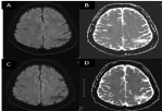
    Figure 1: A and B DWI and ADC sequence showing no diffusion restriction.
C and D follow up imaging showing punctate foci of diffusion restriction in left
ACA territory.
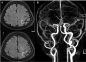
    Figure 2: 2A flair image showing old liosis left posterior parietal region,no
acute infract seen. B: Contrast angiogram circle of wills showing no significant
intracranial stenosis. C: Follow up FLAIR image showing signal changes in
left aca territory (arrows).
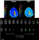
    Figure 3: A and B Perfusion map showing prolonged T max and MIT(Mean
transit time).
Figure 3C: Rapid software showing no diffusion restriction and ischemic
penumbra of 15ml with a mismatch of 15ml.
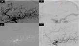
    Figure 4: A Lateral view of cerebral DSAshowing filling defect suggestive of
thrombus in left ACAA2 segment(orange arrow).
B: Delayed Phases showing slow filling internal parietal branch of left
ACA(orange arrows).
C:Roadmap showing microwire in ocduded branch of left Aca(arrows).
Discussion
Imaging forms the backbone of stroke management. Multimodal MRI (DWI,ADC,SWI, FLAIR, MR Angio and perfusion) can provide useful infor¬mation for accurate diagnosis of stroke, evaluation of the risks and benefits of thrombolysis, and prediction of outcomes [8]. Diffusion weighted imaging is a highly sensitive tool for detecting acute ischemia. However it may fail to demonstrate an acute stroke in about 20% cases. Diffusion is the random Brownian motion of the molecules driven by thermal energy. In human body, water is divided between intracellular and extracellular compartments. In an infarcted brain, there is failure of sodium-potassium atpase pump resulting in shift of water from extracellular to intracellular compartment ultimately leading to cell edema and death. This is reflected as diffusion hyperintensity on MRI. Fundamentally four ranges of regional Cerebral Blood Flow (CBF) values quantitatively define normal parenchyma (60–100 mL/100 g/min), oligemic tissue (22–60 mL/100 g/min), penumbra (10–22 mL/100 g/min), and infarction core (< 10 mL/100 g/min)[9]. At CBF levels such as 15 to 20 mL/100 g per minute, infarction may not develop for more than 2 hours [10]. As a result if perfusion defect does not cross a certain threshold level to cause cytotoxic oedema, the DWI images may not show the classical hyperintense lesions seen in acute infarction, though clinical symptoms may be present due to ischemic penumbra. In our patient this could explain the DWI negativity. Also DW-MRI performed early may fail to demonstrate an acute infarct in anterior circulation.
Prevalence of DWI- negative AIS is 5.8% to 9.5% [7,11] (Oppenheim, lian zuo). DWI – negative AIS is now an increasingly recognised entity, and mainly described for posterior circulation strokes [12]. However, only few studies report DWI-negative MRI in anterior circulation stroke [13-16] . Wang et al [13] and Lefkowitz et al [14] reported 2 cases of false negativity involving lesions located in the anterior circulation. They proposed that the degree of hypoperfusion in the early phase of ischemia remained below the threshold required to form an image with DWI as the reason for the false negativity, confirming with our finding. Meta-analysis by Edlow. et.al [12] concluded that causes of DWI negative strokes could be due to small volume of infarct especially in brainstem stroke and hyperacute stroke in which acute ischemia may be underestimated or missed. Bulut et al [17] studied DWI negative strokes and found equal number of cases involving anterior and posterior circulation. They stated the fact that false-negative DWI findings in acute stroke not only involve the posterior circulation and small lacunar or subcortical lesions but that they can also be observed in the anterior circulation and with large lesions.
DW-MRI negativity may have an impact on clinical decisionmaking process in centres using MRI as primary screening tool for diagnosis and to guide therapy in patient with acute stroke. The possibility of false negative DW-MRI in AIS should be kept in mind to avoid misdiagnosis or delay in treatment [7,12]. In our patient MR perfusion study aided in diagnosis of acute ischemia. It determines the ischemic penumbra which is a region of restricted blood supply in which the metabolism is preserved. This ischemic penumbra is vulnerable to progressing to infarcted dead tissue when the delayed perfusion exceeds a certain threshold. According to Lee et el [18], PWI (perfusion weighted imaging) abnormalities, and especially focal perfusion abnormalities, were independently associated with new acute lesions in follow-up DWI. If the hypoperfused state persists after recanalization, tissue damage in the ischemic penumbra seen as DWI-negative but PWI-positive lesion on MRI may evolve into irreversible cell death and damaged tissue, which will manifest as a DWI-positive lesion in the follow-up MRI. MR perfusion studies in addition to DWI MRI images increases sensitivity of diagnosis of AIS to 97.5% [5]. Also, MR perfusion studies aides in decision making for EVT through its DWI-perfusion mismatch indices and newer artificial intelligence softwares like RAPID which quantifies the ischemic penumbra helping in selecting patients for EVT and improve the outcomes in patients [19]. Patient had delayed recovery of right leg weakness after successful radiological treatment of ACA occlusion. This was opined to be due to neuronal stunning [20]. Neuronal stunning leading to delayed recovery can be seen as late as 3 months from the date of treatment.
In conclusion DWI MRI imaging is very sensitive but false negative results can be seen. Sensitivity of diffusion can be increased by Optimisation of diffusion sequence by increasing b value, repeating a corona diffusion, increasing spatial resolution and reducing geometric distortion. However that shouldn’t delay the treatment as diagnosis of ischemic stroke should be balanced between clinical and radiological findings. MR perfusion can aid in diagnosing the proportion of DWI negative AIS facilitating faster treatment with reliably excluding stroke mimics.
References
- Chalela JA, Kidwell CS, Nentwich LM, Luby M, Butman JA, Demchuk AM, et al. Magnetic resonance imaging and computed tomography in emergency assessment of patients with suspected acute stroke: a prospective comparison. Lancet Lond Engl. 2007; 369: 293-8.
- Burke JF, Kerber KA, Iwashyna TJ, Morgenstern LB. Wide variation and rising utilization of stroke magnetic resonance imaging: data from 11 states. Ann Neurol. 2012; 71: 179-85.
- Fiebach JB, Schellinger PD, Jansen O, Meyer M, Wilde P, Bender J, et al. CT and diffusion-weighted MR imaging in randomized order: diffusion-weighted imaging results in higher accuracy and lower interrater variability in the diagnosis of hyperacute ischemic stroke. Stroke. 2002; 33: 2206-10.
- Jauch EC, Saver JL, Adams HP, Bruno A, Connors JJB, Demaerschalk BM, et al. Guidelines for the early management of patients with acute ischemic stroke: a guideline for healthcare professionals from the American Heart Association/American Stroke Association. Stroke. 2013; 44: 870-947.
- Simonsen Claus Z, Madsen Mette H, Schmitz Marie L, Mikkelsen Irene K, Fisher Marc, Andersen Grethe. Sensitivity of Diffusion- and Perfusion- Weighted Imaging for Diagnosing Acute Ischemic Stroke Is 97.5%. Stroke. 2015; 46: 98-101.
- Schellinger Peter D, Thomalla Götz, Fiehler Jens, Köhrmann Martin, Molina Carlos A, Neumann-Haefelin Tobias, et al. MRI-Based and CT-Based Thrombolytic Therapy in Acute Stroke Within and Beyond Established Time Windows. Stroke. 2007; 38: 2640-5.
- Oppenheim C, Stanescu R, Dormont D, Crozier S, Marro B, Samson Y, et al. False-negative Diffusion-weighted MR Findings in Acute Ischemic Stroke. 2000; 8: 1434-40.
- Kim BJ, Kang HU, Kim HJ, Ahn SH, Kim NY, Warach S, Kang DW. Magnetic Resonance Imaging in Acute Ischemic Stroke Treatment. Journal of Stroke 2014; 16: 131-145.
- Yuh WTC, Alexander MD, Ueda T, Maeda M, Taoka T, Yamada K, et al. Revisiting Current Golden Rules in Managing Acute Ischemic Stroke: Evaluation of New Strategies to Further Improve Treatment Selection and Outcome. Am J Roentgenol. 2016; 208: 32-41.
- González RG. Clinical MRI of Acute Ischemic Stroke. J Magn Reson Imaging. 2012; 36: 259-71.
- Zuo L, Zhang Y, Xu X, Li Y, Bao H, Hao J, et al. A retrospective analysis of negative diffusion-weighted image results in patients with acute cerebral infarction. Sci Rep. 2015; 5: 8910.
- Edlow BL, Hurwitz S, Edlow JA. Diagnosis of DWI-negative acute ischemic stroke: A meta-analysis. Neurology. 2017; 89: 256-62.
- Wang PY K, Barker PB, Wityk RJ, Ulug AM. Diffusion-Negative Stroke: A Report of Two Cases. 1999; 10: 1876-1880.
- Lefkowitz D, LaBenz M, Nudo SR, Steg RE, Bertoni JM. Hyperacute Ischemic Stroke Missed by Diffusion-Weighted Imaging. 1999; 20: 1871-1875.
- Tong DC, Yenari MA, Albers GW, Brien M, Marks MP, Moseley ME. Correlation of perfusion- and diffusion-weighted MRI with NIHSS score in acute (<6.5 hour) ischemic stroke. Neurology. 1998 ; 50: 864-70.
- Cho T-H, Hermier M, Alawneh JA, Ritzenthaler T, Desestret V, Østergaard L, et al. Total Mismatch: Negative Diffusion-Weighted Imaging but Extensive Perfusion Defect in Acute Stroke. Stroke. 2009 ; 40: 3400-2.
- Bulut HT, Yildirim A, Ekmekci B, Eskut N, Gunbey HP. False-Negative Diffusion-Weighted Imaging in Acute Stroke and its Frequency in Anterior and Posterior Circulation Ischemia: J Comput Assist Tomogr. 2014; 38: 627-33.
- Lee SH, Nah HW, Kim BJ, et al. Role of Perfusion-Weighted Imaging in a Diffusion-Weighted-Imaging-Negativ Ischemic Attack. J Clin Neurol. 2017; 13: 129-137. doi:10.3988/jcn.2017.13.2.129
- Vyas D, Bohra V, Karan V, Huded V. Rapid processing of perfusion and diffusion for ischemic strokes in the extended time window: An Indian experience. Ann Indian Acad Neurol. 2019; 22: 96.
- Alexandrov Andrei V, Hall Christiana E, Labiche Lise A, Wojner Anne W, Grotta James C. Ischemic Stunning of the Brain. Stroke. 2004; 35: 449-52.