
Case Report
Austin J Cardiovasc Dis Atherosclerosis. 2023; 10(2): 1058.
How Much Attention Should Be Paid to Urinary Tract Infection in a Patient with Congenital Heart Disease?
Dragana D Radoicic¹; Stefan Veljkovic¹; Jovana Lakcevic¹; Ivan Ilic1,2; Slobodan Tomic¹; Aleksandra Nikolic1,2
¹Institute for Cardiovascular Disease’ Dedinje’ Belgrade, School of Medicine, Serbia
²Faculty of Medicine, University of Belgrade, Belgrade, Serbia
*Corresponding author: Dragana D.Radoicic Institut for Cardiovascular Disease Dedinje, Heroja Milana Tepica 1, Belgrade, Serbia. Tel: +381 63 38 37 38 Email: dragana.kastratovic@yahoo.com
Received: November 06, 2023 Accepted: December 06, 2023 Published: December 13, 2023
Abstract
Background: Tetralogy of Fallot (TOF) is the most common cyanotic Congenital Heart Disease (CHD). As therapy becomes increasingly effective, the number of adults with CHD is growing. There are many surgical options to resolve the pulmonary stenosis as a part of TOF, and Pulmonary Valve (PV) replacement with biological prosthesis is one of the solutions. We reported a case of prosthetic valve endocarditis in a patient with TOF.
The Case Report: A patient with weeklong fever, cough, and suspicion of infective endocarditis previously admitted to a local hospital was transferred to Cardiovascular Institute the next day. The pulmonary valve replacement was performed three years previous to his admission to the hospital as well as a complete surgical correction of TOF 22 years before that. Transthoracic Echocardiography (TTE) revealed a large vegetation 27x32mm attached to the leaflet with significantly increased embolic potential and a very high transvalvular gradient across the valve (66/37mmHg). These findings have been confirmed by Trans Esophageal Echocardiography (TEE). Empirical antibiotic therapy was administered. Blood cultures and urine samples, that were taken at the patient’s admission to the Institute, received as negative after two days. However, 4 weeks later, the control TEE examination has shown that the large vegetation was persisted with high embolic risk and clear signs of valve dysfunction. Although this patient already had two midline sternotomy, the Heart team had to decide whether to perform another open-heart surgery with pulmonary valve replacement. Intraoperative findings showed thrombus with vegetation on prosthesis and leaflets, both destroyed by endocarditis. These were sent to microbiological and antibiotic sensitivity analysis and afterward, the valve was replaced with biological prosthesis SJM Supra Epic 27. Cultures of the vegetation identified Staphylococcus epidermitis. Six weeks of antibiotic treatment was applied after the surgical procedure. The patient has been followed up for a year without complications.
Keywords: Congenital heart disease; Pulmonary valve; Infective endocarditis; Staphylococcus epidermitis; Urinary infection
Introduction
Congenital Heart Diseases (CHD) are the most common types of birth defects; with the incidence of 10/1000 live births [1]. The number of adults with some form of CHD is rapidly growing as therapy becomes increasingly effective. It is estimated that 90% of patients with CHD reach adulthood [2]. Tetralogy of Fallot (TOF) as an often cyanotic heart disease can be treated with several different surgical procedures. Treatment of pulmonary stenosis as a part of TOF depends on the stenosis level and localization. Common treatments are valve commissurotomy with a trans-annular patch, pulmonary valve replacement, and conduit. Prosthetic valve dysfunction is frequently a long-term complication after pulmonary valve replacement (age between 15-20 is average) [5]. Patients with CHD are more susceptible to a higher incidence of endocarditis (11 per 100000 persons/year) than the general population therefore it is considered to be the most prevalent underlying condition in patients with Infective Endocarditis (IE) [6]. Inappropriate treatment of urinary infection as a risk factor for IE is associated with serious dysfunction of a prosthetic valve on a pulmonary position and may require open-heart surgery with valve replacement.
The Case Report
A 29 years old male patient with a history of valve replacement caused by TOF was admitted to our tertiary clinic, complaining about weeklong fever, cough, and headache. His vitals parameters revealed very high body temperature (40,6°C) and laboratory findings showed the elevation of WBC 14,6x10³ with 82% polymorphs, anemia with hemoglobin and hematocrit of 11g/dl and 29% respectively. C reactive protein has been proven to be elevated 100,8mg/dl, while procalcitonin had negative value (<0,15ng/ml).
The patient’s anamnesis included surgical correction of TOF at the age of four with resection of the infundibulum and ventricular septal defect patch as well as commissurotomy of the bicuspid pulmonary valve. Owing to inappropriate results, the surgical repair with a transannular patch including the reconstruction of the main pulmonary artery was necessary for the patient. In 2016 the patient underwent bio prosthetic pulmonary valve replacement and reconstruction of truncus pulmonalis.
On a physical exam, he had a regular heart rhythm, with normal first sound and fixed split-second sound, without heart murmur. Electrocardiogram showed a sinus rhythm with an advanced right bundle branch block, a 150 QRS axis and his blood pressure level was normal. Chest CT showed pleural effusion right with compressive atelectasis of the lower part of the right lung.
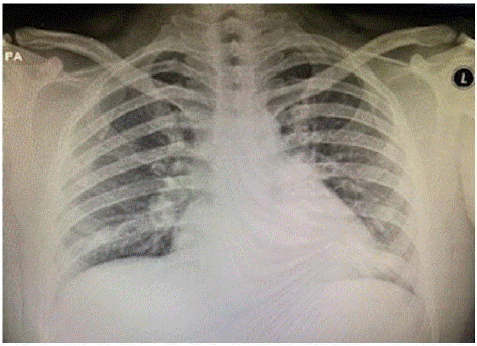
Figure 1: Chest X- ray was performed in lying position and showed small pleural effusion in the lower right side.
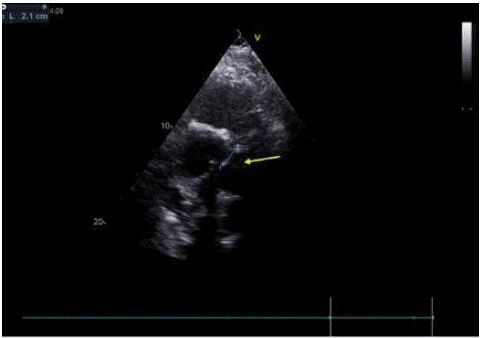
Figure 2: Right Ventricular Inflow Tract (RVIT) reveals a large, highly mobile, echogenic mass (27x23) consistent with a vegetation of PV.
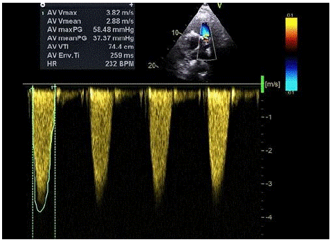
Figure 3: Color Doppler and pressure gradient across the biological valve peak/mean gradient 58/37mmHg.tion of PV.
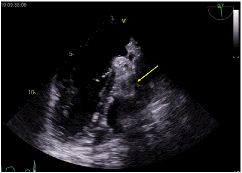
    Figure 4: Transesophageal echocardiographic findings in pulmonary prosthesis infective endocarditis: large vegetation

attached to the pulmonary prosthesis at mid esophageal level.
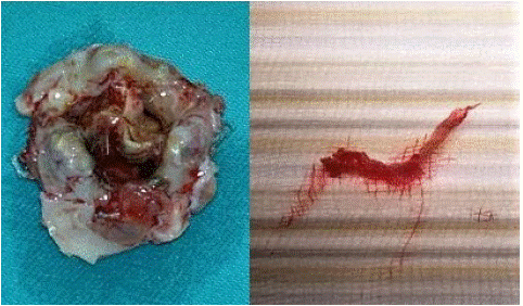
Figure 5: left: Intraoperative finding of filiform vegetation on the thrombus; right: Biological prosthesis destroyed by IE with a native leaflets; right: filiform vegetation.
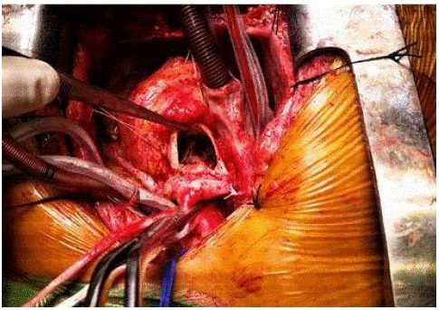
Figure 6: Open-heart surgery.
At admission, a TTE echocardiography was performed showing that a left atrium and a left ventricle were normal sizes with preserved ejection fraction. The right ventricle was dilated, with mild tricuspid regurgitation, and right ventricular systolic pressure was 24mmHg remaining within normal function limits. A biological prosthesis is in the position of the pulmonary valve. An echo-dense filiform mass with dimensions of 27x23mm was attached to the left leaflet. It was mobile, adherent to the valve, and was similar to a large oscillating mass with increased embolic risk [4]. Several smaller masses located at the ventricular side of the biological valve also were reminiscent of vegetation. By using Doppler ultrasound, the peak pressure gradient across the valve was estimated as 66mm Hg while the mean was 41mm Hg. No pulmonary regurgitation was found. Other valves were normal.
Two physicians with extensive experience in echocardiographic examination evaluated TEE. They revealed a highly mobile oscillating mass on the pulmonary artery side, dimension of 27x23mm, with high risk for thromboembolism, as well as a few smaller on the ventricular side, suggesting vegetation presence.
All three sets of blood cultures and samples for urinary analysis were taken while the patient while the patient was febrile at admission in the hospital. Empirical parenteral therapy vancomycin (2g/day) and gentamicin (160mg/day) were administered. Two days after the multiple blood cultures results were sterile and lately, in the blood analyses, c-reactive protein was normalized after 11 days. Results for urine analysis were negative. The patient’s co-existing risk factor for developing infective endocarditis, which he mentioned afterwards, was the presence of clinical symptoms of recurrent urinary infections in the past half-year. He said that he treated himself by using some antibiotics (sulphonamide) for a day or two without doctors’ prescription until the symptoms disappeared.
The Heart team decided to perform a pulmonary valve replacement by open-heart surgery along with an antibiotic treatment during the following 6 weeks based on the findings of the control TEE showing a persisting dimension of vegetations with high transvalvular gradient even after four weeks from the initial examination. Initial vancomycin (2g/day) and gentamicin (160mg/day) therapy was prescribed during the first two weeks and after that period, vancomycin dose was adapted according to its correctly prescribed therapy level in the blood for the next 4 weeks combined with meropenem. Antibiotic therapy was introduced during 4 weeks before surgery as well as during 2 weeks afterwards.
A midline sternotomy with cardiopulmonary bypass was performed and then the RVOT and main pulmonary artery were incised longitudinally. Biological valve Trifecta No 27 was extracted as it was destroyed by endocarditis such as leaflets. Intraoperative finding showed that there was a thrombus with the attached vegetation on the prosthesis. Heart tissue samples were acquired during surgery and forwarded to microbiological and antibiotic sensitivity analysis. Other valves were not affected. The pulmonary valve was replaced by a bio-prosthesis SJM Supra Epic No 27 and then RVOT and truncus pulmonalis by pericardial patch reconstruction was performed.
The vegetation cultures revealed Staphylococcus epidermitis, and susceptibility testing showed sensitivity to complete range of antibiotics that were already applied, accordingly the Heart team decided to complete the treatment after 6 weeks.
During the postoperative period, the patient was afebrile, hemodynamically stable, and discharged from the hospital after the antibiotic therapy was administrated. He was followed up for a year after surgery and maintained satisfactory health condition. The first postoperative echocardiogram in the PV position showed biological prosthesis with a peak/main transvalvular gradient 23/14mmHg.
Discussion
Infective endocarditis of the pulmonary valve is still a rare entity with an incidence of about 1.2% and high mortality and morbidity [8]. The real reasons for such a low prevalence are assumed to be low-pressure gradient to the right heart, the low prevalence of valve disease, and the fact that lower oxygen content of the venous blood in the right chamber may decrease the virulence of aerobic bacteria [3]. The most frequent microbiological findings for PVE are Streptococcus spp. followed by Staphylococcus spp. with methicillin-resistant Staphylococcus aureus and Staphylococcus epidermitis [10]. In our patient’s case, a microbiological agent was isolated from the vegetation on the valve after surgery. The PVE diagnosis was based on the clinical symptoms nature, the blood cultures results, laboratory dates, cardiac imaging, and echocardiography. Blood culture samples were obtained during the patient's febrile condition and after antibiotic administration. Urine analysis was performed using samples obtained immediately upon admission to the hospital as well as during the hospitalization.
Echocardiography is a crucial part of the diagnosis and evaluation of IE. A modified version of the Duke criteria recommends TEE as a preferred modality for the evaluation of PVE. The sensitivity of TEE for the diagnosis of PVE was reported to be around 85-90%, whereas some studies found the sensitivity of TTE to be below 60% [8].
The diagnosis of PVE is still very rare, but there is a need for higher clinical suspicion among patients with CHD in hospitals that are not specialized for valvular heart disease because it can cause serious complications and mortality. According to results of CONCOR ACHD study, it indicates valve-containing prosthetics as a main determinant of IE risk [11]. Current European and American guidelines [12] recommend IE prophylaxis in patients with prosthetic material used for valve replacement or valve repair, and during the first 6 months after implantation of other prosthetics. This case showed how unrecognized and inappropriately treated urinary infections in the past could cause huge dysfunction of the prosthetic valve. Increasing transvalvular gradient for more than 3 times in two years (echocardiography control was performed after the first year only and the gradient across the valve was the same as the one after the surgery was performed 22/13mmHg) and large vegetation that did not react on antibiotic treatment were vital criteria for another surgical treatment with very serious risk. Robinson et al. observed that although 100% of vegetation fewer than 10mm responded to medical therapy alone, only 63% of vegetation over 10mm did, with the rest requiring surgery [9]. Based on our experience, the average lifetime of bioprosthesis ranges between 15 and 20 years. However, in these kinds of cases with such a rapid dysfunction of the valve, surgical procedure is necessary because currently there is not any other quite as effective medicament treatment.
References
- Hoffman JIE, Kaplan S. The incidence of congenital heart disease. J Am Coll Cardiol. 2002; 39: 1890-900.
- Marelli AJ, Mackie AS. Congenital heart disease in the general population circulation AHA. 2007; 106: 627224.
- European journal of echocardiography, recommendations for the practice of echocardiography in infective endocarditis; 2009.
- Gulack C, Ehsan B. Brian, Pulmonary Valve Replacement with a Trifecta Valve Is Associated with Reduced Transvalvular Gradient. Available from: Annalsthoracicsurgery.org.
- Endocarditis in congenital heart disease: CirculationÄHA. 2013; 128: 1396-7.
- Liekiene D, Bezuska L, Semeniene P, Cypiene R, Lebetkevicius V, Tarutis V, et al. Surgical Treatment of Infective Endocarditis in Pulmonary Position -15 years single centre experience. Medicina (Kaunas). 2019; 55: 608.
- Miranda WR, Connolly HM, Bonnichsen CR, DeSimone DC, Dearani JA, Maleszewski JJ, et al. Prosthetic pulmonary valve and pulmonary conduit endocarditis: clinical, microbiological and echocardiographic features in adults. Eur Heart J Cardiovasc Imaging. 2016; 17: 936-43.
- Right-sided valvular endocarditis: etiology, Diagnosis an Approach to Therapy-Robbins et al. 1986; 111: 128-35.
- Japanese National Collaboration. Study. Heart Infect Endocarditis Congenit Heart Dis. 2005; 91: 795-800.
- Kuijepers JM. Koolbergen Dr, Groenink M, Incidence, risk factors and predictors of infective endocarditis in adult congenital heart disease focus on use of prostetic materials. Eur Heart J. 2017; 38: 2048-2056.
- Baddour LM, Walter FR, Wilson etc. infective Endocarditis in Adults: diagnosis, antimicrobial therapy and Menangement of Complication. Circulation. 2015; 132: 1435-86.