
Case Report
Austin Cardio & Cardiovasc Case Rep. 2025; 9(1): 1066.
Cardiac Metastasis of Pulmonary Adenocarcinoma: Case Report
Yassine Alfatihi*
Cardiology Department, Military Hospital of Marrakech, Marrakech, Morocco
*Corresponding author: Yassine Alfatihi, Cardiology Department, Military Hospital of Marrakech, Marrakech, Morocco Tel : 0668913783; Email : dr.yassine.alfatihi@gmail.com
Received: August 19, 2025 Accepted: September 09, 2025 Published: September 12, 2025
Abstract
Secondary cardiac tumors are a serious and relatively rare pathology, typically associated with an advanced stage of the primary tumor and consequently with a poor prognosis. We report the case of a 72-year-old patient followed for pulmonary adenocarcinoma who underwent a chest-abdomenpelvis (CAP) CT scan as part of a staging workup.
Introduction
Lung cancer is the leading cause of cancer-related death in both men and women worldwide. Primary pulmonary carcinoma spreading to pulmonary vessels is relatively rare, and only a few individual cases have been reported in the medical literature [1].
Case Presentation
A previously healthy 72-year-old patient consulted his general practitioner due to general health deterioration and a weight loss of 10 kg over 2 months. He did not report any specific symptoms except for anorexia, exertional dyspnea, and a chronic cough.
His medical history revealed that he had been smoking two packs of cigarettes per day since the age of 30. On physical examination, the patient showed signs of general decline but no dyspnea at rest. Cardiac auscultation was normal. Pulmonary auscultation revealed the absence of breath sounds in the left lung field. A chest CT scan revealed a left pulmonary mass, and the histopathological examination confirmed a pulmonary adenocarcinoma. He was referred to us for a CAP CT scan as part of the staging evaluation.
The CT scan showed a left hilar mass syndrome extending 10 cm anteroposteriorly, (Figure A) crossing the midline with multiple lymphadenopathies near the pulmonary artery trunk, as well as a well-defined, oval mass in the left atrium with the same density as the pulmonary mass, in addition to adrenal and brain involvement.
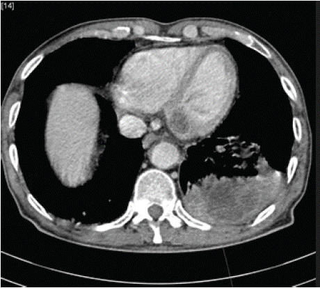
    Figure 1: CT scan showed a left hilar mass syndrome extending 10 cm
anteroposteriorly.
Discussion
Secondary cardiac tumors are rare but serious clinical entities. Malignant tumors can spread to the heart via direct infiltration (figure B), hematogenous metastases, lymphatic metastases, or transvenous extension. For example, renal cell, testicular, and hepatocellular carcinomas can sometimes extend into the inferior vena cava and gradually reach the right atrium. Pulmonary tumors can directly invade the heart through the pulmonary veins. This is a rare occurrence, especially when the lung tumor is small and located far from the heart [3]. Cardiac invasion is generally associated with poor prognosis.
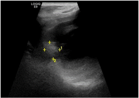
Figure 2: Echocardiography showing a mass in the left atrium.
Clinically, left heart invasion can be life-threatening, leading to complications such as pulmonary venous obstruction [4], cardiac tamponade [5], ventricular arrhythmias [6], complete atrioventricular block [7], left ventricular outflow obstruction [8], and myocardial infarction [9].
Thrombus formation in the pulmonary artery due to direct tumor invasion has also been reported [10]. Anticoagulant therapy is generally the treatment of choice in cases of intracardiac thrombus. Moreover, malignant thrombus in the left superior pulmonary vein with intracardiac extension appears to result from direct intravascular spread of the disease. These patients are at high risk of sudden cardiac arrest due to pulmonary artery obstruction, mitral valve inflow blockage, or massive tumor emboli affecting major organs [11].
In a previous review of 215 lung cancer patients studied with gadolinium-enhanced 3D MR angiography, involvement of the proximal pulmonary veins and left atrial extension was found in 9 (4.2%) and 2 (0.9%) patients, respectively [12]. Similarly, a more recent retrospective analysis of 4668 patients who underwent surgery for lung cancer found pathological signs of pulmonary vein and left atrial involvement in 34 (0.7%) and 25 (0.5%) cases, respectively [13].
Bronchogenic carcinoma develops around the bronchus and may encase pulmonary vessels. As these are low-pressure vascular structures, extrinsic compression and occlusion can occur. This should not be misinterpreted as a vascular "cut-off" due to embolism. The key is a tapered narrowing caused by extrinsic compression, as opposed to a sharp transition typical of embolism.
Tumors can also invade the pulmonary artery. Tumors arising in the pulmonary arteries include angiosarcoma and tumor emboli from other organs. They are a rare but important differential diagnosis of pulmonary embolism [14].
Patients most often present with symptoms related to lung cancer (e.g., cough, hemoptysis, weight loss) or sometimes with cardiac complications at the time of first clinical presentation.
Although patients with cardiac metastases generally have poor clinical outcomes, their management should include a careful evaluation of surgical options. If possible, the treatment of choice is complete surgical resection combined with chemotherapy or radiotherapy [32].
Unfortunately, this was not the case for our patient, who had multiple metastases and thus advanced-stage disease. After thorough clinical evaluation, the patient was deemed to be at too high risk for thoracic or brain surgery. Moreover, complete tumor resection is not always feasible, and postoperative mortality is relatively high. In such cases, cardiac treatment is generally limited to palliative interventions aimed at relieving cardiac compression or hemodynamic obstruction if indicated, along with careful anticoagulant therapy.
Conclusion
Cardiac metastases from pulmonary adenocarcinoma are rare and indicate advanced disease, where the prognosis is poor and treatment is often symptomatic and palliative.
References
- Stella F., Dell’Amore A., Caroli G. Surgical results and long-term follow-up of T(4)-non-small cell lung cancer invading the left atrium or the intrapericardial base of the pulmonary veins. Interact Cardiovasc Thorac Surg. 2012; 14: 415–419.
- Takahashi K., Furuse M., Hanaoka H. Pulmonary vein and left atrial invasion by lung cancer: assessment by breath-hold gadolinium-enhanced threedimensional MR angiography. J Comput Assist Tomogr. 2000; 24: 557–561.
- Dedeilias P., Koletsis E., Rousakis A.G., Kouerinis I., Zaragkas S., Grigorakis A. Deep hypothermia and circulatory arrest in the surgical management of renal tumors with cavoatrial extension. J Card Surg. 2009; 24: 617–623.
- Liaw C.-C., Chang H., Yang T.-S., Wen M.-S. Pulmonary venous obstruction in cancer patients. J Oncol. 2015; 2015: 10.
- Gowda R.M., Khan I.A., Mehta N.J. Cardiac tamponade and superior vena cava syndrome in lung cancer – a case report. Angiology. 2004; 55: 691–695.
- Kinoshita K., Hanibuchi M., Kishi M., Kanematsu T., Nishioka Y., Sone S. Case of squamous cell lung cancer with myocardial metastasis complicated with ventricular tachycardia. J Jpn Respir Soc. 2009; 47: 817–822.
- Morio S., Hara Y., Yamaga T., Yoshino Y., Nakamoto S., Sugiyoma N. A case report of complete heart block by metastatic cardiac involvement from lung cancer. Jpn J Thorac Surg. 1989; 42: 944–947.
- Brandt R.R., Rubin J., Reeder G.S. Intracardiac extension of a lung tumor causing left ventricular inflow obstruction. J Am Soc Echocardiogr. 1995; 8: 930–933.
- Kinjo Y., Nagasaki A., Teruya I. Cardiac involvement of lung cancer presenting with acute myocardial infarction-like electrocardiographic changes. Intern Med. 2006; 45: 113–114.
- Goto T., Maeshima A., Kato R. Lung adenocarcinoma with peculiar growth to the pulmonary artery and thrombus formation: report of a case. World J Surg Oncol. 2012; 21: 10–16.
- Lin M.T., Ku S.C., Wu M.Z., Yu C.J. Intracardiac extension of lung cancer via the pulmonary vein. Thorax. 2008; 63: 1122.
- Takahashi K., Furuse M., Hanaoka H. Pulmonary vein and left atrial invasion by lung cancer: assessment by breath-hold gadolinium-enhanced threedimensional MR angiography. J Comput Assist Tomogr. 2000; 24: 557–561.
- Riquet M., Grand B., Arame A. Lung cancer invading the pericardium: quantum of lymph nodes. Ann Thorac Surg. 2010; 90: 1773–1777.
- Dranoff G. Cytokines in cancer pathogenesis and cancer therapy. Nat Rev Cancer. 2004; 4: 11–22.