
Case Report
Austin Cardio & Cardiovasc Case Rep. 2016;1(2): 1007.
Electrocardiographic Manifestations in Three Psychiatric Patients with Hypothermia – Case Report
Pelechas E¹*, Tsigaridas N², Kyrama S³, Trogganis E4 and Kardamis Ch5
¹Accident and Emergency Department, Scarborough General Hospital, United Kingdom
²Department of Cardiology, Chatzikosta Hospital, Ioannina, Greece
³Department of Cardiology, General Hospital of Arta, Greece
4Department of Cardiology, General Hospital of Kastoria, Greece
5Department of Cardiology, General Hospital of Corfu, Greece
*Corresponding author: Eleftherios Pelechas, Accident and Emergency Department, Scarborough General Hospital, Scarborough, North Yorkshire, United Kingdom
Received: June 02, 2016; Accepted: June 13, 2016; Published: June 15, 2016
Abstract
Hypothermia occurs when the core body temperature falls below 35deg;C and, in severe cases, it can lead to electrocardiographic changes. Several conditions which can occur in the psychiatric population increase the risk of hypothermia which can be aggravated by the use of several classes of medications such as antipsychotics, beta-adrenergic antagonists, benzodiazepines and other sedatives. Three psychiatric patients have been admitted for hypothermia and electrocardiographic manifestations (sinus bradycardia, QT prolongation and Osborn waves) which reversed completely after treatment.
Keywords: Hypothermia; Osborn waves; Electrocardiographic changes; Psychiatric patients.
Introduction
Hypothermia is associated with a spectrum of electrocardiographic changes [1]. The degree of hypothermia leads to various electrocardiographic manifestations [2]. In mild hypothermia (35deg;C - 32deg;C), the Electrocardiogram (ECG) is usually normal but it can rarely show J waves (Osborn waves) [3]. The presence of Osborn waves in inferior and lateral leads, in combination with the appearance of other electrocardiographic manifestations such as increase in PR and QT intervals, increase in QRS complex duration, decrease in amplitude of P and T waves and frequent supraventricular arrhythmias, are noted in moderate hypothermia (32deg;C – 28°C) [4-7]. In severe hypothermia (<28deg;C), additional ECG changes such as J waves in all leads, absence of P waves and frequent ventricular arrhythmias [8-9]. Osborn wave is considered the most specific ECG change in hypothermia [10-12].
Case Presentation
Three psychiatric inmates,within a month (December), have been transferred to the emergency department by ambulance due to low responsiveness (two of them) and coma (the third patient). The medical and drug history of those three patients is presented in (Table 1). Their electrocardiogram showed sinus bradycardia (38bpm – 43bpm), QT prolongation (.52sec - .72sec) and Osborn waves (Figure 1, Figure 2 and Figure 3). There is also a “shivering artifact” on the electrocardiograms of the first and the third patient.
![]()
    Age (in years)
    Drug history
    Core body    temperature


    Case 1
    50
    Diazepam, haloperidol, zolpidem
    27,2deg;C


    Case 2
    61
    Diazepam, haloperidol, zolpidem,
      clozapine, biperiden

      27,6deg;C


    Case 3
    64
    Levodopa, memantine, amlodipine/valsartan
      , lorazepam

      28,8deg;C
Table 1: Medical and drug history of the three cases of hypothermia.
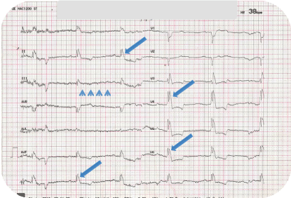
    Figure 1: Electrocardiogram in severe hypothermia (27,2deg;C): sinus
bradycardia (38bpm), QT prolongation, J waves in all leads, shivering artefact.
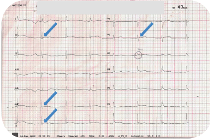
    Figure 2: Electrocardiogram in severe hypothermia (27,6deg;C): sinus
bradycardia (43bpm), QT prolongation, J waves and myocardial infarctionlike
ST elevation in V4 – V6.
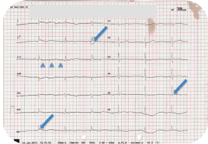
    Figure 3: Electrocardiogram in moderate to severe hypothermia (28,8deg;C):
sinus bradycardia (38bpm), QT prolongation, J waves in inferior leads (II, III,
avf), shivering artefact.
All of those three patients were under treatment with benzodiazepines (diazepam and lorazepam), one of them was also suffering from enuresis and the other two from Parkinson’s disease, which was poor-controlled. Their full blood count, urea and electrolytes and chest x-ray film did not show any signs of infection. Because of the fact that we had three patients in so short period of time, we found out that the heating system was not working as it should and it could be one of the factors as well as the enuresis problems and the shakings of the poor-controlled Parkinson’s disease, that led those patients in hypothermia.
Case nr. 1 was taking orally diazepam and haloperidol when both increase sedation and drowsiness while combining zolpidem and diazepam increases risk of central nervous system depression.
Case nr. 2 was using the same medication with case nr. 1 plus clozapine. Combination of diazepam, haloperidol and clozapine increase sedation and drowsiness and there is risk of heart or respiratory failure.
The only possible interaction in case nr. 3 is that of amlodipine and valsartan with levodopa, which both can produce added drug effects and should be monitored closely.
After the progressive slow rewarming of the patients with special rewarming blankets, the electrocardiographic manifestations came back to normal. They have been discharged a few days later.
Discussion
J wave was initially reported by Kraus 1920 and then in 1922 in a patient with hypercalcemia and later Tomazewski (1938) in a patient with hypothermia [13-15]. John Osborn reported experimentally induced J wave in hypothermic dogs in 1953. He described J wave as a “current of injury” and postulated its occurrence secondary to hypothermia-induced acidosis [16]. Osborn waves (also known as camel-hump sign, late delta wave, hathook junction, hypothermic wave, prominent J wave), are positive deflections occurring at the junction between the QRS complex and the ST segment, where the S point (also known as the J point), has a myocardial infarction-like elevation [17-18]. The mean vector axis of the J wave is oriented anteriorly, inferiorly and leftward across the left ventricle and septum [6,19] J waves are present in 80% of patients with a temperature less than 35deg;C [2]. There is no consensus on the prognostic significance of J waves. The physiological basis of J wave has been described by Antzelevitch and colleagues [19]. Responsible for the characteristic spike and dome pattern of action potential in the ventricular epicardial and endocardial cells is the presence of 4-aminopyridinesensitive transient outward potassium current [20]. This current is more prominent in the ventricular epicardium compared to the endocardium. This difference creates a voltage gradient between the epicardial and endocardial cells [21]. This voltage gradient across ventricular myocardium is accentuated by the hypothermia and this results in prominent Osborn waves.
Osborn waves are notpathognomic of hypothermia. Normothermic patients can also present those waves. Some causes of Osborn waves in normothermic individuals are presented in (Table 2) [22-26]. Although the arrhythmogenic implications of the Osborn waves are not fully understood, the existence of this characteristic deflection may represent some underlying critical conditions. Different medical backgrounds of each patient could lead to ventricular arrhythmias but this should be considered individually. The true significance of the Osborn waves needs further studies in order to be considered as a potential distinguished characteristic for the patients who present it.
![]()
    Acute    ischemic events


    Cocaine    use


    Haloperidol    overdose


    Left    ventricular hypertrophy


    Hypercalcemia


    Brugada’s    syndrome


    Central    nervous system injury


    After    resuscitation of cardiac arrest
Table 2: Causes of Osborn waves in normothermic patients.
Several conditionsthat can occur in the psychiatric population, increase the risk of hypothermia. Mental retardation, debilitating physical illness, nocturnal enuresis and seizure disorders are some of those. This risk can be further increased by the use of several classes of medications used to treat psychiatric disorders such as benzodiazepines and other sedatives, antipsychotics, beta adrenergic antagonists [27-28]. Air-conditioning or poor supervision of those patients, regarding the room temperature, can lead to hypothermia.
References
- Gavaliatis IP. Electrocardiographic issues related to action potential phases 1 and 2 on the occasion of a case of accidental mild hypothermia. Int J Cardiol. 2001; 77: 81-86.
- Cheng D. The EKG of hypothermia. J Emerg Med. 2002; 22: 87-91.
- Aslam AF, Aslam AK, Vasavada BC, Khan IA. Hypothermia: Evaluation, Electrocardiographic Manifestations, and Management. Am J Med. 2006; 119: 297-301.
- Anand K, Radhakrishnan S, Radhakrishnan A. The First Case Report of Accidental Severe Hypothermia from Tropical South India.World J Med. 2014; 10: 446-451.
- Schmidt-Schweda S, Ohler A, Post H, Pieske B. Moderate hypothermia for severe cardiogenic shock (COOL Shock Study I & II). Resuscitation. 2013; 84: 319-325.
- Mustafa S, Shaikh N, Gowda RM, Khan IA. Electrocardiographic features of hypothermia. Cardiology. 2005; 103: 118-119.
- Mattu A, Brady WJ, Perron AD. Electrocardiographic manifestations of hypothermia. Am J Emerg Med. 2002; 20: 314-326.
- Ansari E, Cook JR. Profound hypothermia mimicking a Brugada type ECG. J Electrocardiol. 2003; 36: 257-260.
- Graham CA, McNaughton GW, Wyatt JP. The electrocardiogram in hypothermia. Wilderness Environ Med. 2001; 12: 232-235.
- Junttila MJ, Sager SJ, Tikkanen JT, Anttonen O, Huikuri HV, Myerburg RJ . Clinical significance of variants of J-points and J-waves: early repolarization patterns and risk. Eur Heart J. 2012; 33: 2639-2643.
- Antzelevitch C, Yan GX. J wave syndromes. Heart Rhythm. 2010; 7: 549-558.
- Edelman ER, Joynt K. J waves of Osborn revisited. J Am Coll Cardiol. 2010; 55: 2287.
- Kraus F. UeberdieWirkung des Kalziums auf den Kreislauf. Dtsch Med Wochensch. 1920; 46: 201-203.
- Kraus F, Zondek SG. Uber die Durchtrankungsspannung. Klin Wochensch I Jahrgang. 1922; 36: 1778-1779.
- Tomaszewski W. Changements electrocardiographiques observes chez un homme mort de froid. Arch Mal Coer. 1938; 31: 525.
- Osborn JJ. Experimental hypothermia: Respiratory and blood pH changes in relation to cardiac function. Am J Physiol. 1953; 175: 389-398.
- Maruyama M, Kobayashi Y, Kodani E, Hirayama Y, Atarashi H, Katoh T, et al. Osborn waves: history and significance. Indian Pacing Electrophysiol J. 2004; 4: 33-39.
- Gussak I, Bjerregaard P, Egan TM, Chaitman BR. ECG phenomenon called the J wave. History, pathophysiology, and clinical significance. J Electrocardiol. 1995; 28: 49-58.
- Yan GX, Antzelevitch C. Cellular basis for the electrocardiographic J wave. Circulation. 1996; 93: 372-379.
- Antzelevitch C, Sicouri S, Litovsky SH, Lukas A, Krishnan SC, Di Diego JM, et al. Heterogeneity within the ventricular wall. Electrophysiology and pharmacology of epicardial, endocardial, and M cells. Circ Res. 1991; 69: 1427-1449.
- Litovsky SH, Antzelevitch C. Transient outward current prominent in canine ventricular epicardium but not endocardium. Circ Res. 1988; 62: 116-126.
- Patel A, Getsos JP, Moussa G, Damato AN. The Osborn wave of hypothermia in normothermic patients. Clin Cardiol. 1994; 17: 273-276.
- Otero J, Lenihan DJ. The “normothermic” Osborn wave induced by severe hypercalcemia. Tex Heart Inst J. 2000; 27: 316-317.
- Alings M, Wilde A. Brugada’s syndrome: clinical data and suggested pathophysiological mechanism. Circulation 1999; 99: 666-673.
- De Sweit J. Changes simulating hypothermia in the electrocardiogram in subarachnoid hemorrhage. J Electrocardiol. 1972; 5: 93-95.
- Jain U, Wallis DE, Shah K, Blakeman BM, Moran JF. Electrocardiographic J waves after resuscitation from cardiac arrest. Chest. 1990; 98: 1294-1296.
- van Marum RJ, Wegewijs MA, Loonen AJ, Beers E . Hypothermia following antipsychotic drug use. Eur J Clin Pharmacol. 2007; 63: 627-631.
- Hägg S, Mjörndal T, Lindqvist L. Repeated episodes of hypothermia in a subject treated with haloperidol, levomepromazine, olanzapine, and thioridazine. J Clin Psychopharmacol. 2001; 21: 113-115.