
Case Report
J Blood Disord. 2023; 10(2): 1078.
Intravascular Large B-Cell Lymphoma Detected by Cutaneous Biopsy with Pancytopenia and Negative Bone Marrow Biopsy
Battis N¹*; Gardeen S²; Kozlowski A²; Furda-Raine L¹
¹Department of Dermatology, Park Nicollet Health System, USA
²Department of Dermatology, Health Partners Institute, USA
*Corresponding author: Battis N Department of Dermatology, Park Nicollet Health System, 7550 34th Ave. S., Suite 101, Minneapolis, MN 55450, USA. Tel: (+1) 952-220-1064; Fax: 952-883-9746 Email: nicholas.battis@parknicollet.com
Received: June 23, 2023 Accepted: July 25, 2023 Published: August 01, 2023
Abstract
Intravascular large B-cell lymphoma is a B-cell lineage neoplasm, which is typically recognized as a high-grade neoplasm with poor outcomes. Though rare, the cutaneous variant can present with multiple lesions of the skin and negative systemic staging. We present a case of intravascular large B-cell lymphoma first identified on cutaneous biopsy, with pancytopenia, in addition to negative bone marrow biopsy and serum protein electrophoresis. The patient is a 71-year-old male who presented with upper body pruritis and pancytopenia, as well as weight loss and fatigue. A full body skin exam incidentally revealed a well-demarcated, firm, bound down atrophic lichenified plaque on the right anterior thigh. A punch biopsy revealed large, atypical lymphoid cells exclusively localized within the lumina of variably sized blood vessels, immunoreactive for CD20, BCL6, MUM1, and CD5, but negative for CD3, CD10, cyclin D1, and TdT. A diagnosis of intravascular large B-cell lymphoma was made, and he was referred to oncology for further treatment. Full body skin exam is a mainstay of dermatologic practice, and it is essential to pursue biopsy with uncharacteristic lesions, regardless of the presenting concern.
Keywords: IVLBCL; Lymphoma; Intravascular; Cutaneous biopsy; Intravascular large B-cell lymphoma
Abbreviations
CRP: C-Reactive Protein; RBC: Red Blood Cell; WBC: White Blood Cell; PLT: Platelet; SPEP: Serum Protein Electrophoresis; IVLBCL: Intravascular Large B-cell Lymphoma; DIF: Direct Immunofluorescence; FISH: Fluorescence in Situ Hybridization; HLH: Hemophagocytic Lymphohistiocytosis; R-HD-MTX-ARA-C: Rituximab with High-Dose Methotrexate and Cytarabine; R-CHOP: Rituximab, Cyclophosphamide, Doxorubicin Hydrochloride, Vincristine, and Prednisone; WHO: World Health Organization
Case Presentation
We present a case of intravascular large B-cell lymphoma first identified on cutaneous biopsy, with pancytopenia, in addition to negative bone marrow biopsy and serum protein electrophoresis.
A 71-year-old male presented to dermatology clinic on referral from oncology due upper body pruritus with pancytopenia, weight loss, and fatigue. On exam, he had well-defined, eroded, and crusted papules in various stages of healing on the bilateral arms and upper back with ill-defined, subtle, thin, pink papules coalescing into thin plaques on the neck. On the right anterior thigh, a well-demarcated, firm, bound down atrophic lichenified plaque was incidentally noted. The patient was adamant that this was due to crossing his legs while watching television, however when having the patient cross his legs, there was no appreciable pressure in the location of the plaque.
Laboratory evaluation was notable for an elevated Anti-Nuclear Antibody (ANA) of 1:640, copper of 153.4 μg/dL, ferritin of 2,124 ng/mL, and C-Reactive Protein (CRP) of 4.1 mg/dL. Also notable was his pancytopenia with a Red Blood Cell (RBC) count of 3.18*1012/L, White Blood Cell (WBC) count of 3.4*109/L, and Platelet (PLT) count of 55*109/L.
Prior to presentation in dermatology, a bone marrow biopsy was hypocellular without increase in blasts, nor morphologic evidence of lymphoma. Serum Protein Electrophoresis (SPEP) was within normal limits. Given his presentation without unifying diagnosis, skin biopsies were performed (Figure 1).
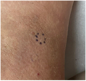
Figure 1: Atrophic, bound down, lichenified plaque noted on exam. The lesion measured approximately 5 cm in diameter. The site of acquired biopsy is encircled.
A telescoping punch biopsy of the anterior right thigh revealed large, atypical lymphoid cells exclusively localized within the lumina of variably sized blood vessels (Figure 2), consistent with Intravascular Large B-Cell Lymphoma (IVLBCL). Lesional cells were immunoreactive for CD20, BCL6, MUM1, and CD5, but negative for CD3, CD10, cyclin D1, and TdT (Figure 3). Direct Immunofluorescence (DIF) showed granular deposition of both IgM and C3 along the basement membrane zone. Fluorescence in situ hybridization (FISH) showed no MYC (8q) or MYC/IGH (8;14) rearrangement.
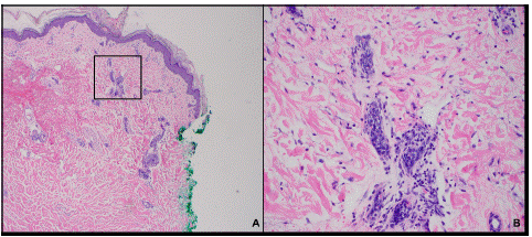
Figure 2: Hematoxylin and Eosin (H&E) stain of anterior right thigh punch biopsy at 40x (A) and 100x (B) magnification. Atypical lymphoid cells can be seen exclusively localized within the lumina of variably sized blood vessels. The section of the slide seen in image B is outlined in black on image A.
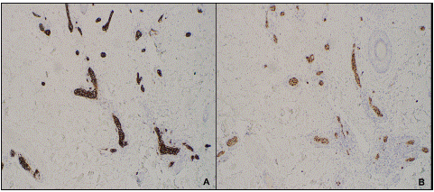
Figure 3: Immunostaining positive for CD20 (A) and MUM1 (B). Stains are visualized at 100x magnification.
A diagnosis of IVLBCL, with cutaneous and Hemophagocytic Lymphohistiocytosis (HLH) variants was made. He was initiated on rituximab with high-dose methotrexate and cytarabine (R-HD-MTX-ARA-C), alternating with rituximab, cyclophosphamide, doxorubicin hydrochloride, vincristine, and prednisone (R-CHOP) per oncology.
Pancytopenia trended toward normal and at 2-month dermatologic follow-up, there was significant symptomatic improvement of his pruritus. The lichenified patch on the right medial thigh showed marked reduction in size and induration. He continues to follow with oncology and dermatology at 3-month intervals.
Discussion
IVLBCL is a B-cell lineage neoplasm, which is typically recognized as a high-grade neoplasm with poor outcomes [1]. This lymphoma is defined by the World Health Organization (WHO) as a B-cell lymphoma showing predominant intravascular growth [2]. Neoplastic cells do not commonly circulate into the peripheral blood or form extravascular masses, leading to frequently missed or post-mortem diagnoses [1,3]. The classical variant of IVLBCL presents with nonspecific B signs, such as fever of unknown origin, however cutaneous variants have been described [3-4].
The cutaneous variant typically presents with multiple lesions of the skin and negative systemic staging [4]. Typically, laboratory results demonstrate normal leukocyte and platelet counts, which contrasts with what was seen in our patient. The cutaneous variant of IVLBCL has been most frequently reported in western nations, though more recent information has also shown increasing prevalence of the neurological subtype in these geographic regions [4,5]. The cutaneous variant of IVLBCL shows a strong predilection for females, as demonstrated in a study which found that 100% (n=10) of reported cases were seen in female patients [4].
In consideration of our patient’s normalizing blood counts and lack of systemic symptoms aside from generalized pruritis, cutaneous variant IVLBCL carries a more favorable prognosis. When compared with classical variant IVLBCL, the cutaneous variant demonstrates an improved 3-year overall survival [56%±16% vs 22%±10%] [4].
Though the cutaneous variant is less aggressive, treatment is the same as classical IVLBCL and must be treated as a disseminated disease. R-CHOP, as used in our patient, has high efficacy against both the cutaneous and classical variants, with 90% of patients showing complete remission and 2-year disease free progression in 56% of patients [6,7].
Conclusion
This case is unique with consideration to the presentation of IVLBCL only on cutaneous biopsy, with accompanying pancytopenia, despite negative bone marrow biopsy and SPEP. This case demonstrates the importance of full body skin exam in the setting of generalized dermatologic symptoms and pursuing biopsy with uncharacteristic lesions, despite red herring history.
References
- Davis JW, Auerbach A, Crothers BA, Lewin E, Lynch DT, et al. Intravascular large B-cell lymphoma. Arch Pathol Lab Med. 2022; 146: 1160-7.
- Swerdlow SH, Campo E, Harris NL, et al, editors. WHO classification of tumours of haematopoietic and lymphoid tissues. rev 4th ed. Lyon, France: IARC. World Health Organization Classification of Tumours. 2017; 2.
- Ponzoni M, Campo E, Nakamura S. Intravascular large B-cell lymphoma: a chameleon with multiple faces and many masks. Blood. 2018; 132: 1561-7.
- Ferreri AJ, Campo E, Seymour JF, Willemze R, Ilariucci F, et al. Intravascular lymphoma: clinical presentation, natural history, management and prognostic factors in a series of 38 cases, with special emphasis on the ’cutaneous variant’. Br J Haematol. 2004; 127: 173-83.
- Brunet V, Marouan S, Routy JP, Hashem MA, Bernier V, et al. Retrospective study of intravascular large B-cell lymphoma cases diagnosed in Quebec: A retrospective study of 29 case reports. Med (Baltim). 2017; 96: e5985.
- Ferreri AJ, Dognini GP, Bairey O, Szomor A, Montalbán C, et al. The addition of rituximab to anthracycline-based chemotherapy significantly improves outcome in ’Western’ patients with intravascular large B-cell lymphoma. Br J Haematol. 2008; 143: 253-7.
- Shimada K, Matsue K, Yamamoto K, Murase T, Ichikawa N, et al. Retrospective analysis of intravascular large B-cell lymphoma treated with rituximab-containing chemotherapy as reported by the IVL study group in Japan. J Clin Oncol. 2008; 26: 3189-95.