
Case Report
Austin Biomark Diagn. 2016; 3(1): 1023.
Vertebral Artery Dissection Associated with Bathing: A Case Report
Xingyong Chen, Xu Zhang, Xiaosong Wang, Huixin Lei and Yinzhou Wang*
Department of Neurology, Fujian Medical University Shengli Clinical College, PR China
*Corresponding author: Yinzhou Wang, Department of Neurology, Fujian Provincial Hospital, Fujian Medical University Shengli Clinical College, PR China
Received: September 30, 2015; Accepted: January 21, 2016; Published: January 22, 2016
Abstract
Vertebral Artery Dissection (VAD) is often associated with trauma or occurs spontaneously, inevitably causing some neurological deficits. We herein report the case of a 49-year-old female patient presented with sudden onset of severe posterior neck pain with occipital headache, vertigo and vomiting when she was bathing. Dissection of the right vertebral artery at the level of V4 segment was confirmed by use of Magnetic Resonance Imaging (MRI) and CT Angiography (CTA), and Digital Subtraction Angiography (DSA). The patient was started with clopidogrel (75mg/d) and low molecular weight heparin calcium injection (Fraxiparine, 0.4ml: 4100AXaIU/bid, subcutaneous injection). Recovery of neurologic function was excellent, and she was discharged after 21 days. However, reexamine CTA showed an abnormal intracranial segment of the right vertebral artery. The patient still continued treatment with Clopidogrel (75mg/d). We suggest that patients with symptoms of neck pain or headache and even disturbances of posterior circulation after manipulation and stretching of the neck, VAD has to be taken into consideration.
Keywords: Vertebral artery dissection; Magnetic resonance; CT Angiography; Digital subtraction angiography
Introduction
Spontaneous dissection of the intracranial vertebral artery is an increasingly recognized cause of stroke. The annual incidence of spontaneous Vertebral Artery Dissection (VAD) is 1-1.5 per 100,000 [1]. Usually, a history of drug treatment, generalized convulsive seizure, minor blunt trauma or activity associated with rotation or hyperextension of the neck, such as tooth-brushing, aerobics, yoga, painting of the ceiling and spinal manipulations can cause VAD [2- 5], and it is generally labeled as spontaneous dissection. The purpose of this case report is to describe a female patient who presented with posterior neck and occipital pain and was undergoing a vertebral artery dissection when she was standing in a shower and washing her hair. The VAD caused embolic complications: pontine and cerebellar infarctions.
Case Presentation
A 49-year-old female patient presented with sudden onset of severe posterior neck pain with occipital headache, vertigo and vomiting when she was bathing two hours ago. When she was admitted to our department, she appeared tired and distressed. She stated that she had burning, sharp pain worse than she had ever experienced before. She was healthy previously, and denied any head or neck trauma and chiropractic manipulation. She has no personal or family history of systemic illnesses, connective tissue diseases, myocardial infarctions or cerebrovascular insults. On physical examination, her temperature was 37.60C, pulse 72 beats/min, and blood pressure 132/78 mmHg. No remarkable positive signs were detected by the lung, cardiac, and abdominal examinations. Neurological examination revealed a right-sided slight hemiparesis with Babinski sign, neck stiffness, a right-sided central facial palsy and an ataxic finger-nose test as well as heel-knee-shin on the bilateral side. In addition, the blood samples for kidney and liver function, blood and urine routine examination, coagulation factors and antithrombin III were all in the normal range of reference. The electrocardiogram and chest X-ray was normal. Brain Magnetic Resonance Imaging (MRI) scans showed an acute infarct involving the bilateral cerebella (A) hemisphere and a leftsided pontine (B) infarction (Figure 1). CT Angiography (CTA) of the neck and head revealed dissection of right distal vertebral artery (Figure 2). Moreover, Digital Subtraction Angiography (DSA) showed focal luminal dilatation involving right V4 segment of the vertebral artery just proximal to Posterior Inferior Cerebellar Artery (PICA) and insufficient blood flow to its distal portion (Figure 3). Dissection of the right vertebral artery at the level of V4 segment was confirmed. The patient was started with clopidogrel (75mg/d) and low molecular weight heparin calcium injection (Fraxiparine, 0.4ml: 4100AXaIU/ bid, subcutaneous injection). Recovery of neurologic function was excellent, and she was discharged after 21 days. However, reexamine CTA after 20 days showed an abnormal intracranial segment of the right vertebral artery (Figure 4). The patient still continued treatment with clopidogrel (75mg/d).
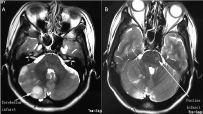
    Figure 1:  MRI T2-weighted axial images showing the bilateral cerebellar (A)
and pontine (B) infarcts (arrow).
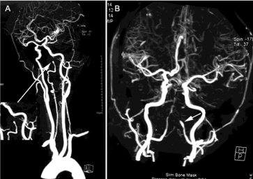
    Figure 2:  CTA of Craniocervical arteries showing proximal vessel lumen
segmental expansion and distal lumen flame like tapering of the right
intracranial vertebral artery near to baseline artery (arrow).
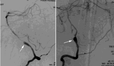
    Figure 3:  DSA showed an abnormal vertebral artery segmental lumen
expansion and distal lumen slender of the right intracranial vertebral artery
near to baseline artery (arrow).
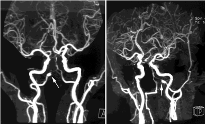
    Figure 4:  Reexamine CTA after 20 days showed an abnormal intracranial
segment of the right vertebral artery (arrow).
Discussion
VAD recently has been recognized as an important cause of ischemic stroke in middle-aged adults (10-25% of these cases) [6]. The anatomy of the vertebral arteries (ascendance parallel to the spine through the transverse foramina of the C1-C6) makes them vulnerable to stretching and compression of the spine, especially at the C1/ C2 levels, where arteries are relatively fixed and where a big part of head rotation occurs [2]. In particular, the neck region is vulnerable, where hyperextension, rotation, flexion and distraction movements can occur, which are known to cause VAD [7,8]. Given the mundane nature of these activities and the fact that the vast majority of the population practices them without ever developing VAD (a rare condition), it is unlikely that they could be considered a “cause” of the condition [9]. We suggested that there may be no causal relationship and that general forces of manipulation may not be enough to cause VAD. It is more likely that previous damage or a preexisting defect was present and that an otherwise trivial trauma triggered the VAD [10]. In this case, we postulated the patient maybe present these factors include connective tissue disorders, hypertension, recent infection, vessel abnormalities, and atherosclerosis. Vertebral artery dissection is the result of a flap-like tear in the tunica intima of the vertebral artery. Because of this tear, blood enters into the tunica media causing a hematoma in the vessel wall. The development of intramural hematoma due to the dissection resulted in narrow lumen, pseudoaneurysm and hypoperfusion of vertebrobasilar system. Usually, intracranial vertebral artery dissection has two major types of presentation: focal neurological deficits due to vertebrobasilar artery ischemia and subarachnoid hemorrhage. About 60% of VAD patients have symptoms of vertebrobasilar circulation ischemia, such as vertigo, hoarseness, dysphagia and cerebellar dysfunction [11]. Occipital headache and neck pain are common symptoms and occur in 70% of patients with VAD [12]. The acute onset of posterior neck and occipital headache in our patient was thought to result from VAD. Since ipsilateral PICA is usually not well compensated, thus infarction of right lateral cerebellar hemisphere mostly developed, and the patient experienced ataxia and hemiparesis of right face and right extremities. The other possible mechanism might be related to transient embolization of vertebrobasilar system from the VAD, or related to a combination of embolization and local occlusion [13].
Accurate diagnosis and appropriate management of VAD depend on the knowledge of its clinical course and serial radiographic features. MRI and MRA are providing a noninvasive method for evaluating arterial dissection. In this case, initial suspicion of vertebral artery dissection was raised by MR findings, it was reported that MRI and MRA had a sensitivity of 20% and a specificity of 100% for vertebral artery dissection [14], which prompted us to perform cerebral angiography for definitive diagnosis. CTA data can be viewed as source images, maximum-intensity-projection images, volume-rendered (3D reconstruction) images, and multiplanar reformation images. DSA is more sensitive than these other tests for subtle dissections [14]. Similarly, when discrepant or equivocal results are seen on CTA, at our institution, we may favor DSA as the most suitable next study. It may be asked under what circumstances DSA should be performed for dissection, when noninvasive tests without patient risk are available. The first circumstance involves cases in which CTA, MR techniques, or both provide discrepant results [14]. The second circumstance involves acute clinical situations in which, if a dissection is found, an endovascular procedure (e.g., intra-arterial stent placement) will likely be needed soon afterward. In such cases, in the interest of time, DSA could reasonably be deemed the most appropriate diagnostic study. A meta-analysis showed that different endovascular treatment modalities are comparatively safe and effective in the management of vertebral artery dissection [15]. Their reduced operative time, minimal invasiveness, and overall safety render them a suitable option for intervention-amenable dissections [15]. However, we preferred to antithrombotic with clopidogrel and low molecular weight heparin calcium injection (Fraxiparine) rather than endovascular interventional treatment in this patient since the latter strategy might lead to greater risks and disadvantages.
The final outcome of VAD is related with the clinical and angiographic characteristics of the disease. Intracranial vertebral artery dissection with subarachnoid hemorrhage is notorious for frequent rebleeding and a poor prognosis [16]. Nevertheless, some patients survive with a good final outcome. The factors associated with the prognosis of this disease are not fully understood and appropriate treatment strategies continue to be debated [16]. It was reported that poor admission neurological grade, rebleeding episode(s), and lesions with a pearl-and-string structure were predictive of poor outcomes [16]. Those patients with these characteristics may be candidates for aggressive attempts to prevent rebleeding during the acute stage. Endovascular internal trapping for VAD is a therapy with a satisfactory long-term outcome [17]. Conversely, patients without these characteristics may be good candidates for conservative treatment, especially those who survive the acute phase without rebleeding [16]. The long-term clinical outcomes for symptomatic intracranial unruptured vertebrobasilar artery dissection (siu-VBD) were favorable in all patients without ischemic symptoms and in most patients with ischemic presentation [18]. None of the siu-VBD caused subarachnoid hemorrhage. Old age and basilar artery involvement were independent predictors of unfavorable outcome in siu-VBD with ischemic presentation [18]. So far, our patient was favorable and was still in follow-up.
Conclusion
In conclusion, VAD is an important cause of stroke in the young. Neck pain or headache is an important warning symptom of VAD. In patients with symptoms of disturbances of posterior circulation after manipulation and stretching of the neck, VAD has to be taken into consideration. MRA and CTA should be firstly performed for discovering the dissection. DSA could reasonably be deemed the most appropriate diagnostic study. Early recognition of VAD is paramount, as timely initiation of appropriate anticoagulation can be crucial in ensuring full recovery.
References
- Redekop GJ. Extracranial carotid and vertebral artery dissection: a review. Can J Neurol Sci. 2008; 35: 146-152.
- Amin FM, Larsen VA, Tfelt-Hansen P. Vertebral artery dissection associated with generalized convulsive seizures: a case report. Case Rep Neurol. 2013; 5: 125-129.
- Mosby JS, Duray SM. Vertebral artery dissection in a patient practicing self-manipulation of the neck. J Chiropr Med. 2011; 10: 283-287.
- Shi S, Chen K, Ge X, Ni B. Lessons from the diagnosis and treatment of spontaneous vertebral arterial dissection. Case report. Interv Neuroradiol. 2009; 15: 203-208.
- Mantia-Smaldone GM, Bagley LJ, Kasner SE, Chu CS. Vertebral artery dissection and cerebral infarction in a patient with recurrent ovarian cancer receiving bevacizumab. Gynecol Oncol Case Rep. 2013; 5: 37-39.
- Schievink WI. Spontaneous dissection of the carotid and vertebral arteries. N Engl J Med. 2001; 344: 898-906.
- Albuquerque FC, Hu YC, Dashti SR, Abla AA, Clark JC, Alkire B, et al. Craniocervical arterial dissections as sequelae of chiropractic manipulation: patterns of injury and management. J Neurosurg. 2011; 115: 1197-1205.
- Reuter U, Hamling M, Kavuk I, Einhaupl KM, Schielke E. Vertebral artery dissections after chiropractic neck manipulation in Germany over three years. J Neurol. 2006; 253: 724-730.
- Mosby JS, Duray SM. Vertebral artery dissection in a patient practicing self-manipulation of the neck. J Chiropr Med. 2011; 10: 283-287.
- Wuest S, Symons B, Leonard T, Herzog W. Preliminary report: biomechanics of vertebral artery segments C1-C6 during cervical spinal manipulation. J Manipulative Physiol Ther. 2010; 33: 273-278.
- Kratz SN, Butler KH. Vertebral artery dissection presenting as acute cerebrovascular accident. J Emerg Med. 2011; 40: 151-157.
- Mokri B, Houser OW, Sandok BA, Piepgras DG. Spontaneous dissections of the vertebral arteries. Neurology. 1988; 38: 880-885.
- Thanvi B, Munshi SK, Dawson SL, Robinson TG. Carotid and vertebral artery dissection syndromes. Postgrad Med J. 2005; 81: 383-388.
- Provenzale JM, Sarikaya B. Comparison of test performance characteristics of MRI, MR angiography, and CT angiography in the diagnosis of carotid and vertebral artery dissection: a review of the medical literature. AJR Am J Roentgenol. 2009; 193: 1167-1174.
- Hernandez-Duran S, Ogilvy CS. Clinical outcomes of patients with vertebral artery dissection treated endovascularly: a meta-analysis. Neurosurg Rev. 2014; 37: 569-577.
- Yamada M, Kitahara T, Kurata A, Fujii K, Miyasaka Y. Intracranial vertebral artery dissection with subarachnoid hemorrhage: clinical characteristics and outcomes in conservatively treated patients. J Neurosurg. 2004; 101: 25-30.
- Kashiwazaki D, Ushikoshi S, Asano T, Kuroda S, Houkin K. Long-term clinical and radiological results of endovascular internal trapping in vertebral artery dissection. Neuroradiology. 2013; 55: 201-206.
- Kim BM, Kim SH, Kim DI, Shin YS, Suh SH, Kim DJ, et al. Outcomes and prognostic factors of intracranial unruptured vertebrobasilar artery dissection. Neurology. 2011; 76: 1735-1741.