
Case Report
J Bacteriol Mycol. 2018; 5(7): 1084.
Clinical Case of Leishmaniosis and Hemophagocytic Syndrome
Gambino F*¹, Martorana G², Rizzo A², Cottone M²
¹Department of Internal Medicine, University of Palermo, Italy
²Biomedical Department of Internal and Specialized Medicine (DI.BI.MIS), University of Palermo, Italy
*Corresponding author: Fabrizio Gambino, Biomedical Department of Internal and Specialized Medicine (DI.BI.MIS), University of Palermo, Italy
Received: October 20, 2018; Accepted: November 06, 2018; Published: November 13, 2018
Case Report
A 72 years old Italian man who had a one-month journey in the South of India in June 2015, was admitted at the emergency room of V. Cervello Hospital in Palermo in July 2017 with a 2 years-old history of erythema nodosum, purple cutaneous nodules, pancytopenia, splenomegaly, and recurrent pulmonary thromboembolism (treated with NOAC and heparin). The patient was admitted two times in two different hospitals where an extensive research for possible infectious (virologic, bacterial and mycological), immunological (all patterns of autoantibodies, complement) and hematological (bone marrow biopsy, Jack2 mutation) causes was performed without any positive result. A diagnosis of suspected vasculitis was performed in the second hospital despite no evidence of histological significative findings and negativity of ANCA. For this reason a steroid therapy was started.
At admission the patient was in poor general conditions, in unconscious state, with precarious vital parameters, fever (present from 15 days) and dyspnoeic. Physical examination showed a purple cutaneous nodule on the anterior surface of the right arm, widespread ecchymosis and hematomas, and a trophic ulcer on the anterior surface of the right leg.
Laboratory values showed partial respiratory failure (low pO2 and normal PCO2), pancytopenia (Hb 9,7 g/dl [normal range 13-17 g/dL], PLT 32000 cells/Ul [normal range 150000-400000 cells/uL], WBC 1221cells/uL, with neutrophils 838cells/uL [normal range WBC 4000-10000 cells/uL-neutrophils 2000-8000 cells/uL], elevated LDH: 1455U/L [normal range: 50-150 U/L], hypertriglyceridemia and hyperferritinemia; an abdominal ultrasound exam showed moderate splenomegaly (largest dimension: 18cm). According to the validated diagnostic criteria, we suspected the haemophagocytic syndrome [as known as Hemophagocytic lymphohistiocytosis [HLH]) and started treatment with Betamethasone, Cyclosporin and Filgrastim [1]. The patient was also treated with broad-spectrum antibiotics and Fluconazole. We performed serological tests and PCR assays for Herpesviridae, HIV, Haepatitis viruses, Widal Wright serodiagnosis, and complete cultures exams screening. PCR for HSV1 DNA and IgM titres against the same virus were positive and so we added Acyclovir to the therapy. Furthermore a chest CT scan showed a massive pulmonary embolism which was treated with Heparin.
Despite the specific therapy for HLH and quick defervescence and moderate clinical improvement, persistent pancytopenia justified a new bone marrow biopsy [2-4], there was a 40% hematopoietic cellularity and copious round bodies into histiocytes-like cells cytosol, morphologically compatible with Leishmanial amastigotes (Figure 1). Splenomegaly, hypergamma-globulinaemia and high IgG titre against Leishmania (1:320) confirmed the diagnosis of Visceral Leishmaniasis.
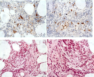
Figure 1: Patient was healthy at follow up 2 year later.
So an Amphotericin-B based therapy (4mg/Kg/die for five continuous days, then weekly-9 doses/total), at the beginning in addition to Cyclosporin and Filgrastim, then without them, was administered [5]. There was a quick healing of the cutaneous ulcer on the leg, of the nodule on the arm and of the widespread ecchymosis and hematomas. The week after we completed Amphoterin-B treatment, negative PCR for leishmanial DNA confirmed the completed recovery from the Leishmanial Infestation. We also completed heparin-based therapy (on the basis of submassive bilateral pulmonary embolism, CT-documented) and antibiotic therapy (for an urinary tract infection), and finally stopped immunosuppressant and steroid (after almost one year and a half of continuous therapy).
References
- Marom D, Offer I, Tamary H, Jaffe CL and Garty. Hemophagocytic Lymphohistiocytosis Associated With Visceral Leishmaniasis. Pediatric Hematology and Oncology. 2009; 18: 65-70.
- Özdemir N, Koç B, Arslanta§ E, Odaman Al I, Kelleci Ç, Uysalol EP and et al. Hemophagocytic Lymphohistiocytosis Associated With Visceral Leishmaniasis. J Pediatr Hematol Oncol. 2018; 40: 395.
- Matnani R, Ganapathi KA. Hemophagocytic lymphohistiocytosis associated with visceral leishmaniasis. Blood. 2016; 127: 513.
- Freeman HR, Ramanan AV. Review of haemophagocytic lymphohistiocytosis. Arch Dis Child. 2011; 96: 688-693.
- Billiau AD, Roskams T, Damme-Lombaerts RV, Matthys P, Wouters C. Macrophage activation syndrome: Characteristic findings on liver biopsy illustrating the key role of activated, IFN-g-producing lymphocytes and IL-6- and TNF-a-producing macrophages. Blood. 2005; 105: 1648-1651.