Case Report
Austin Pathol. 2019; 2(1): 1009.
Unusual Location of a Parasitosis
Damiri A1,2*, Chahdi Ouazzani H1,2, Setti K1,2 and Oukabli M1,2
1Department of Anatomy and Pathology Cytology. Military Hospital Mohammed V Rabat Morocco
2Faculty of Medicine and Pharmacy of Rabat (FMPR), Mohamed V Rabat University, Morocco
*Corresponding author: Damiri A, Department of Anatomy and Pathology Cytology. Mohamed V Rabat University, Morocco
Received: July 12, 2019; Accepted: September 06, 2019; Published: September 13, 2019
Abstract
Oxyuriasis is an intestinal parasitosis, frequent, strictly human, caused by a cosmopolitan roundworm: Enterobius vermicularis. Pinworms are mainly localized in intestine but can sometimes be found in the female genitalia, even if it is rare.
Our case is a young man of 32 who has an anterior history of inconstant anal pruritus but and who presented a pruriginous and painless subcutaneous nodule and who benefited from an excisional biopsy whose pathological examination revealed the presence of sections of nematodes corresponding to female pinworms within a fibro-fatty tissue.
Oxyuriasis is mainly intestinal, sometimes with an uro-genital location, especially in women, but no case has been described at the subcutaneous level, hence the interest of this work in revealing new possible locations of this parasitosis.
Keywords: Pathology, Oxyuriasis, Subcutaneous, Flubendazol
Abbreviations
HE: Hemalin Eosine; G: magnification
Introduction
Oxyuriasis is an intestinal parasitosis, frequent, strictly human, caused by a cosmopolitan roundworm: Enterobius vermicularis. The pinworms are mainly localized in the intestine but can sometimes be found in the female genitals, but their location in the labia minora is much rarer [1].
Adult male and female worms live in the colon. Before dying, fertilized females migrate at night to the anus to lay an average of 10,000 eggs. The eggs thus deposited take 4 to 6 hours to become infectious. They are then transferred to clothing, bedding and dust. Eggs can survive for 15 to 20 days in the environment. Most often, they are transported by the hands (they are mostly found under the nails) and then ingested by the person already infected (selfinoculation) or by a different host.
The ingested eggs hatch in the stomach under the influence of digestive juices and release larvae that migrate to the small intestine. In the intestine, the larvae moult to become adult in 5 to 6 weeks. Worms live about 1 month in the intestine.
Case Presentation
Our patient is a 32-year-old man with no specific history other than an inconstant anal pruritus and who discovered the presence of a subcutaneous nodule at the sacral region, pruritic and painless measuring 1.5 cm and which does not evolve with time. The patient decided to consult a dermatologist who prescribed a dermocorticoid for 1 month without any improvement. After a consultation with a surgeon, a decision to make surgical resection of the nodule was made.
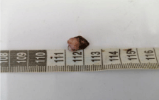
We received in our laboratory a fragment of 1.2x1 cm of yellowish color and soft consistency that was cut in half and included entirely in a block (Figure 1).

    Figure 1: Macroscopic image of the subcutaneous nodule of the sacral
region.
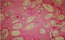
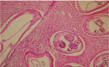
After formalin fixation and paraffin inclusion, histological examination showed fibrofatty tissue with sections of nematodes corresponding to female pinworms containing eggs (Figures 2 and 3). A diagnosis of subcutaneous Oxyuriasis was made.

    Figure 2: Histological image showing fibrous tissue with sections of
nematodes (HE; Gx 10).

    Figure 3: Histological image showing female pinworms containing eggs (HE;
Gx 20).
Discussion
Oxyuriasis occurs all over the world, especially regions with temperate climates, and affects all classes of the population. This is the most common worm infection in the United States; however, according to some studies, its prevalence appears to be decreasing [2].
The prevalence of infection is highest among children between 5 and 14 years. Followed by preschoolers, their family members or caregivers [2].
Infection is easily transmitted within a family (risk of transmission› 75%) and is common in places where children live, play and sleep together (eg day care). otherwise, the risk is lower in schools (risk of transmission ‹10%) [2].
The presence of a big quantity of infectious eggs in the perianal area promotes self-inoculation, especially in individuals putting fingers in mouths. Due to the short lifetime of worms, chronic infections are usually due to repeated reinfections [3].
Although the infection is most often asymptomatic and does not cause any serious medical problems, Oxyuriasis causes anal pruritus, especially at night, irritability, restless sleep and, more rarely, vulva pruritus. Rarely, worms can be observed in feces [3].
Several clinical manifestations, such as teeth grinding, weight loss, and enuresis, have been associated with Oxyuriasis, but no causal relationship has been established.
Transmission is done by eggs ingestion:
- By direct contact.
- By indirect contact: hands, clothing, bedding, towels and washcloths, toilet seats and baths.
- By common vehicle: food.
- By auto-inoculation.
Incubation period:
The incubation period (from eggs ingestion to presence of worms in the perianal region) is 1 to 2 months.
Period of contagiousness:
The contagious period lasts as long as the infected person is untreated and the pregnant females lay their eggs in the perianal area.
Receptivity:
Everyone is susceptible to have an infection caused by pinworms.
Immunity:
Oxyurose does not confer immunity. There is no vaccine against Oxyuriasis.
Oxyurose treatment:
Flubendazole
• If low infestation: 1 tablet or 1 measuring spoon (100 mg tab, suspension of 20 mg/mL),
• If important infestation: 1 tablet morning and evening for 3 days;
or
Albendazole, 1 tablet or 1 measuring spoon (400 mg tab, or 4% oral susp).
Whatever the product, it is essential to combine hygiene measures:
• Change underwear, bedding and towels,
• Cut the nails as close as possible and wash hands before meals,
• Treat all family members the same day,
• Repeat a therapeutic cure 20 days later.
But it happens that pinworms are found in ectopic locations as in the genitals but no case has been published in the literature mentioning a subcutaneous location as the case of our observation.
In fact, female pinworms come to lay in the anal margin and can migrate to the nearby genital organs or to the sacral region as our case.
They can be found in the vagina of women [4, 5], where they can go up the genital tract to end up in the uterus, fallopian tubes, ovaries and in the peritoneum [6]. In these unusual locations, pinworms often cause inflammatory reaction [7], which may result in cervical cancer [8], or even an abscess [9].
A location in Bartholin’s glands has already been reported [10]. In all these cases, a disappearance of the inflammatory phenomena was obtained by the adapted antihelminthic treatment.
Conclusion
Pinworms are intestinal parasites that can sometimes be found in unusual locations [1]. They are then asymptomatic and found fortuitously during an etiological research of another affection [1]. Subcutaneous location remains exceptional and never described in the literature, which raises the question of the route of dissemination in this situation. The treatment remains identical to that of intestinal infestations.
Ethics Approval and Consent to Participate
This work has respected all the rools of medical ethics and has been elaborated by all the authors.
Availability of Material and Data
All data is available in the Military hospital Mohammed V, Rabat, Morocco.
Funding
This work was not funded by a third party payer.
Consent to Publish
As the main author and the names of all authors, I allow you to publish this article in your review
Competing Interests
The authors do not declare any conflict of interest.
Author’s Contributions
References
- Localisation intra-vulvaire de l’oxyurose P. Bourée, F. Plantier, R. Mitri 4 REVUE FRANCOPHONE DES LABORATOIRES • N° 503. 2018.
- Association Française des Enseignants de Parasitologie et Mycologie (ANOFEL) 2014© UMVF - Université Médicale Virtuelle Francopho.
- Maladies infectieuses oxyurose (entérobiase). 2016.
- Chung DI, Kong HH, Yu HS, Kim J, Cho CR. Live female Enterobius vermicularis in the posterior fornix of the vagina of a Korean woman. Korean J Parasitol. 1997; 35: 67-69.
- Kashyap B, Samantray JC, Kumar S, Jhamb R, Singh AK, Kaur IR. Recurrent paediatric pinworm infection of the vagina as a potential reservoir for Enterobius vermicularis. J Helminthol. 2014; 88: 3813.
- Siochou A, Birtsou H, Papazahariadou M. Enterobius vermicularis infection of female genital tract. Int J Immunopathol Pharmacol. 2008; 21: 1031-1033.
- Knuth KR, Fraiz J, Fisch JA, Draper TW. Pinworm infestation of the genital tract. Am Fam Physician. 1988; 38: 127-130.
- Raju K, Verappa S, Venkataramappa SM. Enterobius vermicularis infestation nmasquerading as cervical carcinoma: A cytological diagnosis. J Nat Sci Biol Med. 2015; 6: 476-479.
- Smolyakov R, Talalay B, Yanai-Inbar I, Pak I, Alkan M. Enterobius vermicularis infection of female genital tract: a report of three cases and review of literature. Eur J Obstet Gynecol Reprod Biol. 2003; 107: 220-222.
- Donmez ME, Ozlu T, Yilmaz T, Ayaz E, Enterobius vermicularis: Can it be a possible pathogen in Bartholin gland abscess formation? J Obstet Gynaecol Res. 2014; 4: 268-270.