Introduction
Lichen planus is a chronic inflammatory condition that can affect the skin, mucous membranes, hair, and nails. Its overall prevalence varies from 0.1% to 4% of the global population. Mucosal lichen planus primarily affects the oral mucosa, followed by the genital mucosa, esophagus, and nasopharynx in decreasing order of frequency, with a well-established clinical appearance and histological features that aid in diagnosis [1].
Ocular involvement is a rare occurrence, mainly documented through clinical cases. It can affect various components of the eye, giving rise to a diverse and nonspecific clinical presentation. Symptoms may encompass a spectrum from redness, irritation, and visual disturbances to conditions such as conjunctivitis, blepharitis, and corneal issues. This diversity in symptoms often contributes to diagnostic delays, ultimately leading to chronic and irreversible alterations in the ocular surface's integrity, and thus significant morbidity.
Case Report
We report a case of an 88-year-old patient who was referred from the department of ophthalmology for evaluation of chronic recurrent blepharitis of unknown origin, refractory to multiple topical and oral treatments (including antibiotics, corticosteroids, and cyclines). A medical history review revealed diabetes and hypertension, as well as bilateral cataract surgery six years ago. The patient reported ocular pain, hyperemia, and decline in visual acuity.
The ophthalmologic examination revealed in the right eye a visual acuity of 1/10 with severe blepharitis, symblepharon formation, conjunctival hyperemia with epitheliopathy, corneal neovascularization, and secondary cataract (Figure 1 & 2). In the left eye, visual acuity was limited to counting fingers at a distance, with blepharitis, conjunctival fornix obliteration with symblepharon, conjunctival hyperemia, and axial corneal dystrophy, making it difficult to assess the remainder of the eye (Figure 3 and 4). An exhaustive dermatological examination revealed actinic cheilitis without involvement of the skin, oral mucosa, genital mucosa, or phanerous.
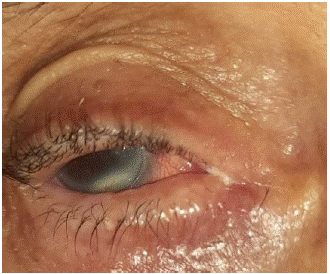
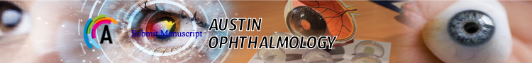
Figure 1: Aspect of blepharitis in the right eye.
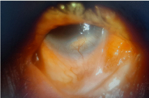
Figure 2: Symblepharons, epitheliopathy, and corneal neovascularization of the right eye.
Figure 3: Aspect of blepharitis in the left eye.
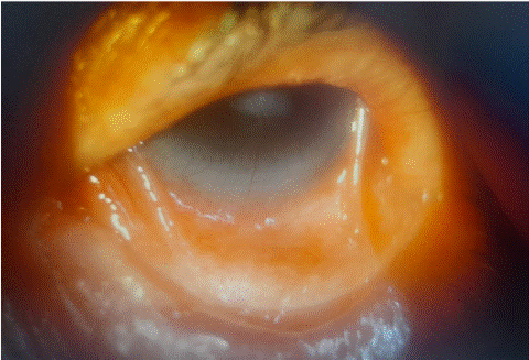
Figure 4: Symblepharons, beginning of deformation of the anatomy of the eyelids and its adnexa of the left eye.
A conjunctival biopsy was performed, revealing a lymphocytic infiltrate with fibrous and hemorrhagic reorganization, and vacuolization of the basal consistent with ocular lichen planus. Indirect immunofluorescence testing for anti-basement membrane antibodies returned negative results. The diagnosis of ocular lichen planus was established, and the patient was started on topical cyclosporine 2% drops treatments with partial improvement.
Discussion
Our case illustrates an isolated and rare occurrence of ocular lichen planus in an 88-year-old patient. It is a rare inflammatory disease characterized by persistent changes in the integrity of the ocular surface. The pathogenetic mechanisms of lichen planus remain elusive; however, prevailing evidence suggests that damage to the epithelium arises from T–cell–mediated keratinocyte apoptosis triggered by an unidentified antigen in genetically predisposed individuals. Various factors, including viruses, autoimmune phenomena, drugs, vaccines, and contact allergens, have been proposed as potential etiological contributors [2].
Ocular lichen planus can affect all components of the eye: eyelids, conjunctiva, anterior and posterior segments, as well as the lacrimal duct. Various clinical aspects have been documented since the early nineteenth century when the first case with conjunctival involvement was reported [3]. This results in a range of manifestations, including conjunctivitis, blepharitis, epitheliopathy, symblepharon, occlusion of the lacrimal duct and corneal perforation, ultimately leading to irreversible damage and blindness. However, these manifestations are not specific to lichen planus, making it challenging to clinically distinguish from other forms of cicatrizing ocular conditions, especially when not associated with other skin or mucosal involvement.
Since its first documentation, approximately 42 cases of ocular lichen planus have been reported [4]. Among these cases, only 11 presented with isolated ocular involvement, while the rest had at least two affected sites, which facilitated their diagnosis. Our patient, who had isolated ocular involvement, brings the total to 12. The patient exhibited manifestations such as blepharitis, symblepharon formation, conjunctival hyperemia with epitheliopathy, and corneal neovascularization, initially misdiagnosed and treated as rosacea.
Ocular lichen planus poses a significant challenge in differential diagnosis, particularly when patients present with cicatricial conjunctivitis. Given that numerous diseases, such as mucous membrane pemphigoid, pemphigus vulgaris, Stevens-Johnson syndrome, and paraneoplastic pemphigus, can manifest with similar clinical features [2] (Table 1), it is crucial that, in cases of cicatricial conjunctivitis or any unexplained ocular symptoms, physicians consider ocular lichen planus among the potential differential diagnoses.
![]()
Causes of Cicatriciel Conjonctivitis
Autoimmune:
Mucous membrane pemphigoid
Linear IgA disease
Lichen planus
Pemphigus vulgaris
Paraneoplastic pemphigusAllergic:
Stevens-Johnson Syndrome
Drug-induced cicatrizing conjunctivitisDiverse
Rosacea
Chemical thermal burns
Neoplasia
Table 1:
A conjunctival biopsy plays a pivotal role in accurately diagnosing the underlying etiology. It reveals lichenoid infiltrates within a thickened basement membrane, irregular acanthosis, basal cell vacuolation, and Civatte bodies. However, these findings are not exclusive to lichen planus. Therefore, Direct Immunofluorescence (DIF) remains valuable in such cases. DIF demonstrates anti-C3 staining in the basement membrane zone and positive anti-IgM staining of Civatte bodies in the papillary dermis. In our case, direct immunofluorescence was not conducted due to the extensive damage to the ocular surface [5].
Treatment strategies encompass both topical and systemic medications to address inflammation and manage specific symptoms. Depending on the severity and extent of ocular involvement, therapies such as cyclosporine, corticosteroids, and other immunomodulatory agents are employed. Topical corticosteroids and cyclosporine are generally considered first-line treatments, with cyclosporine acting to inhibit T lymphocyte proliferation through the inhibition of interleukin-2 receptor expression. Several clinical studies have shown positive outcomes with topical ciclosporin in treating ulcerative or erosive oral lichen planus [6]. In our case, topical cyclosporine was utilized, resulting in partial improvement. Moreover, for more severe or resistant cases, systemic immunosuppressants like mycophenolate mofetil can be considered. However, the rarity of this condition contributes to a lack of substantial evidence. Surgical procedures, such as lacrimal canal drainage, may be suggested in instances of canalicular stenosis and membrane transplantation in case of corneal perofration [7], although the outcomes tend to be modest.
Conclusion
In summary, the occurrence of isolated conjunctival lichen planus is a rare condition that should be considered among the array of diseases that can lead to irreversible scarring keratoconjunctivitis. Confirming a precise diagnosis requires clinical suspicion, substantiated by the execution of a conjunctival biopsy for histopathological and immunofluorescence analysis. There is a heightened importance for both dermatologists and ophthalmologists to be cognizant of this uncommon diagnosis. This awareness is crucial to initiate early and aggressive anti-inflammatory treatment, thereby mitigating the risk of irreversible visual impairment.
References
- Mohebbi M, Mirghorbani M, Banafshe Afshan A, Towfighi M. Lichen Planus in Ocular Surface: Major Presentations and Treatments. Ocul Immunol Inflamm. 2019; 27: 987-94.
- Rozas Muñoz E, Martínez-Escala ME, Juanpere N, Armentia J, Pujol RM, Herrero-González JE. Isolated conjunctival lichen planus: a diagnostic challenge. Arch Dermatol. 2011; 147: 465-7.
- Gaucher Druelle. Lichen planus avec lesions des ongles et localisation sur la conjonction palpebrale Lichen planus with lesions of the nails and localization on the palpebral conjunction. Bull Soc Franc Derm Syph. 1904; 15: 33.
- Safadi M, Viglione M, Zahner S. Ocular lichen planus: An unusual presentation, its treatment, and review of the literature. JAAD Case Rep. 2021; 14: 4-6.
- Neumann R, Dutt CJ, Foster CS. Immunohistopathologic features and therapy of conjunctival lichen planus. Am J Ophthalmol. 1993; 115: 494-500.
- Pakravan M, Klesert TR, Akpek EK. Isolated lichen planus of the conjunctiva. Br J Ophthalmol. 2006; 90: 1325-6.
- Daniela Gomez-Elizondo, Mariana Lopez-Martinez, Raul E Ruiz-Lozano, Jorge E Valdez-García, Julio C Hernandez-Camarena. Corneal perforation associated with isolated ocular lichen planus: a case report. European Journal of Ophthalmology. 2021; 31: NP9-NP12.