
Clinical Image
We report the case of a 75-year-old patient with known hypertensive well followed, monophthalmus of the right eye who consulted for a decrease in visual acuity of the latter. Its best correction is 1/10 at the OD level and negative light perception at the OG level. Eye tone was normal. Biomicroscopic examination revealed central Bietti's dystrophy affecting the lower axis and a grade 1 dense nuclear subcapsular cataract (Figure 1) in the right eye with conjunctivalisation of the right cornea and neovessels (Figure 2). The fundus is inaccessible in both eyes. Ocular ultrasound did not show any retinal detachment. The patient underwent cataract surgery by phacoemulsification and corneal scarification in the right eye which took place without complications with a simple postoperative course and a 4/10 recovery with correction.
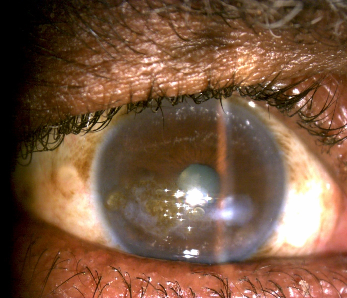
Figure 1:
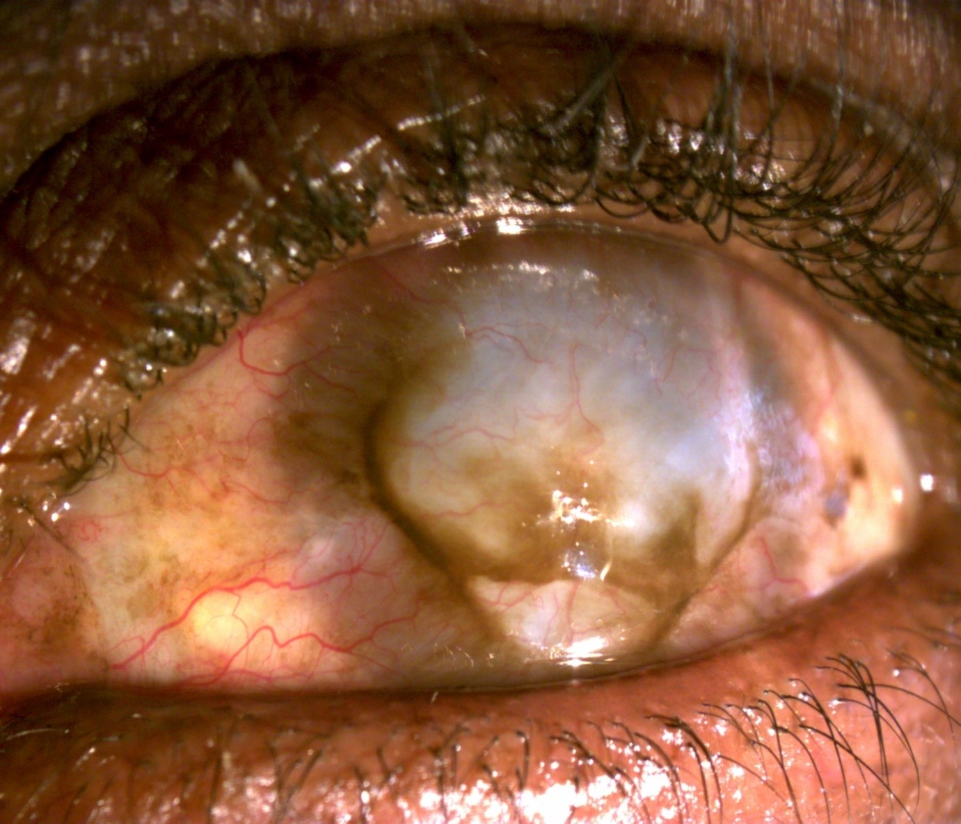
Figure 2: