
Abstract
Craniopharyngiom as are rare epithelial tumors, arising from the pituitary gland or pituitary gland and developing in the sellar and supra sellar region. Their diagnosis is often late and is based on the triad of neurological, ophthalmological and endocrine signs. Their treatment is multidisciplinary and includes surgical and hormonal treatment. Their prognosis depends on the precocity of the diagnosis, the location and the size of the tumor.
Keywords: Craniopharyngioma; Papilledema; Begnin tumor; Cranial hypertension; Surgical resection; Bleomycin
Introduction
Craniopharyngioma in children is a rare benign epithelial tumor, of embryonic origin, it affects both children and adults [1], their incidence is estimated to be 0.5 to 2 new cases per year and per million inhabitants [2], Their treatment is based on surgery completed or not by a hormonal treatment [3].
Case Presentation
We report the case of a 9-year-old child, without any notable pathological history, who presented to the ophthalmologic emergency room for a bilateral visual acuity decrease with a divergent strabismus installed over 2 weeks associated with a paresis of the right lower limb, a urinary leakage with vomiting and an alteration of the general state preceded 2 months earlier by intense and rebellious headaches.
The ophthalmological examination noted the best visual acuity was to 2/10 in the right eye and counting fingers in the left eye, an anisocoria, after dilatation, the fundus examination demonstrated papilledemastage 2, peri-macular exudates (Figures 1 & 2), the examination of the oculomotor nerves showed an incomplete paralysis of the extrinsic 3. In front of this picture a neuro-pediatric examination was performed showing an intracranial hypertension syndrome and an opto-chiasmatic syndrome, completed by a cerebral Magnetic Resonance Imaging (MRI) showing a sellar and supra sellar process evoking a craniopharyngioma with an active left hydrocephalus (Figure 4).
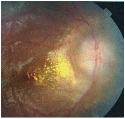
Figure 1: Papilledema with macular exudates in right eye.
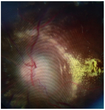
Figure 2: Papilledema with macular exudates in left eye.
Fluorescein angiography was done showing a papillary diffusion of fluorescein confirming the papillary edema of stasis (Figure 3), a visual field was also done agonized.
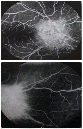
Figure 3: Fluorescein angiography with a papillary diffusion of fluoresce in confirming the papillary edema of stasis.
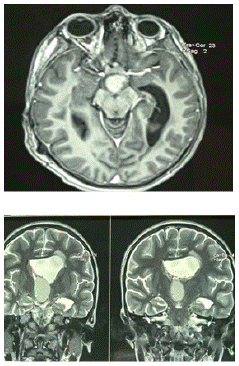
Figure 4: Cerebral IRM showing a craniopharyngioma with an active left hydrocephalus.
The child was referred to the neurosurgery department for rapid decompression with surgical removal.
The rest of the check-ups including hormonal assessment were normal, and the histopathological examination confirmed that the tumor was a craniopharyngioma.
The evolution was marked by a recurrence of the tumor which prompted a treatment with intra-tumoral bleomycin, the evolution was marked by a regression of the tumor with a control at 1 year and 2 years without abnormalities.
References
- Müller HL, Emser A, Faldum A, Bruhnken G, Etavard-Gorris N, et al. Longitudinal study on growth and body mass index before and after diagnosis of childhood craniopharyngioma. J Clin Endocrinol Metab. 2004; 89: 3298-305.
- Van Effenterre R, Boch A.-L, Les craniopharyngiomes. 2007; 826; 399-478. http://dx.doi.org/10.1016/j.ando.2007.08.001
- Puget S, Garnett M, Wray A, Grill J, Habrand JL, et al. Pediatric craniopharyngiomas: classification and treatment according to the degree of hypothalamic involvement. J Neurosurg. 2007; 106: 3-12.
- Elowe-Gruau E, Beltrand J, Brauner R, Pinto G, Samara-Boustani D, et al. Childhood craniopharyngioma: hypothalamus-sparing surgery decreases the risk of obesity. J Clin Endocrinol Metab. 2013; 98: 2376-82.
- Sklar CA. Craniopharyngioma: endocrine abnormalities at presentation. Pediatr Neurosurg. 1994; 21: 18-20.
- Puget S. Treatment strategies in childhood craniopharyngioma. Front Endocrinol (Lausanne). 2012; 3: 64.
- Müller HL. Childhood craniopharyngioma: treatment strategies and outcomes. Expert Rev Neurother. 2014; 14: 187-97.
- Hoffmann A, Postma FP, Sterkenburg AS, Gebhardt U, Muller HL, et al. Eating behavior, weight problems and eating disorders in 101 long-term survivors of childhood-onset craniopharyngioma. J Pediatr Endocrinol Metab. 2015; 28: 35-43.