Clinical Image
Peripheral Ossifying Fibroma (POF) is most often a gingival nodule that is believed to be a reactive lesion rather than a neoplastic pathologic process, with overlapping clinical and histopathological presentations [1]. It is a pedunculated or sessile nodule that presents as a painless, hemorrhagic, and often lobulated mass of gingiva or alveolar mucosa, with area of surface ulcerations [2]. There is variability in the size of the lesion depending upon amount of superficial inflammation and edema [2]. The lesion is typically self-limiting, commonly seen on interdental papilla and comprises about 9% of all gingival growths [3]. Females are more commonly affected, and anterior maxilla is the most prevalent location [1]. POFs are generally diagnosed by clinical inspection and biopsy [3]. Here we present a case of POF with focus on the clinical as well as histological presentation, to explore the possibility for accurate diagnosis.

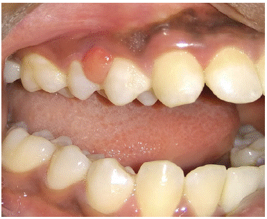
A 20-year-old female patient was reported to the department of Periodontology with the chief complaint of slow growing painless growth that has been present buccally with upper right premoloar region. Growth was associated with bleeding and pain while brushing. The patient did not give any history of trauma, no significant medical and dental history. Intraoral examination revealed a nodular, sessile growth present on the interdental papilla between 14 and 15, extending mesiodistally it was of the same color of the adjacent gingiva with the upper half being more reddish in color. The growth was circular in shape and approximately 5×5 mm in dimensions with well-defined borders. On palpation, it was firm to hard in consistency and non tender. Bleeding on probing was noted. The lesion was asymptomatic, and showed no clinical evidence of ulceration. Based on these findings a provisional diagnosis of pyogenic granuloma was made. Further a differential diagnosis of peripheral ossifying fibroma, peripheral giant cell granuloma was considered. Later with the patients consent an excisional biopsy was performed under aseptic precautions and under local anaesthesia.

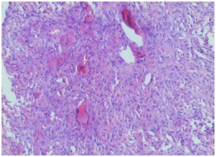
Histopathological features with varying mesenchymal presentations of peripheral ossifying fibroma exhibiting; fasciculate pattern - fibrous proliferation with giant cells The histopathological examination of the lesion revealed hyperplastic stratified squamous epithelium with elongated reteridges with underlying fibrovascular connective tissue. The connective tissue showed rich vascularity with loosely arranged collagen fibers, osteoid like areas and areas showing spicules of bone with osteocytes in the lacunae along with osteoblastic rimming. Chronic inflammatory cell infiltrate consisting predominantly of lymphocytes were seen within the connective tissue with numerous proliferating capillaries along with extravasated RBC. These features confirmed the diagnosis of peripheral ossifying fibroma.
References
- Mergoni G, Meleti M, Magnolo S, Giovannacci I, Corcione L, Vescovi P. Peripheral ossifying fibroma: A clinicopathologic study of 27 cases and review of the literature with emphasis on histomorphologic features. J Indian Soc Periodontol. 2015; 19: 83-7.
- Farquhar T, Maclellan J, Dyment H, Anderson RD. Peripheral ossifying fibroma: a case report. J Can Dent Assoc. 2008; 74: 809-12.
- Ogbureke EI, Vigneswaran N, Seals M, Frey G, Johnson CD, Ogbureke KU. A peripheral giant cell granuloma with extensive osseous metaplasia or a hybrid peripheral giant cell granuloma- peripheral ossifying fibroma: a case report. J Med Case Rep. 2015; 9: 14.
